|

|
|
|
11.2 Deviating chromosome number or structure
|
|
|
|
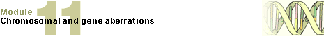
In a reciprocal translocation two broken off chromosome pieces of non-homologous chromosomes are exchanged. This is a relatively frequent anomaly. One finds it with an incidence of 1:500 newborns.
|
|
|
| Reciprocal translocations are frequently balanced because the entire genetic material is present. Problems occur, though, in gamete formation. |
|
Fig. 15 - Reciprocal translocation |
|
Legend |
|

A
B
|
Two non-homologous
chromosome pairs
Reciprocal translocation |
|
|
|
Fig. 15
Two broken off chromosome pieces from various chromosomes grow onto the broken end of another chromosome.
Animation (59 Kb)
|
|
More info
|
|
With reciprocal translocations gamete formation is connected with disorders because chromosomes in the region of the translocation cannot readily pair . In this segment no homologues of the chromosomes are present. For this reason, the involved chromosomes in the reciprocal translocation are forced to put themselves on one another in another form. Quadriradial structures are formed.
More details
|
|
|
|
|
|
The fact that translocations occur repeatedly between the two same chromosome segments on various chromosomes says that DNA sequence homologies exist between such chromosome regions that make a healing process of such pieces easier.
Today it is known that balanced translocations can also lead to pathogenic disorders in that proto-oncogenes, which as normal genes in their customary environment are frequently responsible for the controlling cell proliferation, can be transformed into oncogenes through translocation events (connections with other genes). They are the cause for the origin of many tumors and types of cancer because in other environments they achieve totally different effects. An example for this is the reciprocal translocation between the long arms of chromosomes 9 and 22. Through this, out of a proto-oncogene, arises an oncogene that is responsible for the uncontrolled proliferation of leukocytes in chronic myeloid leukemia (CML).
|
|
|
|
Robertsonian translocation
|
|
|
Another frequently observed anomaly (1:1'000 newborns) is the robertsonian translocation, which occurs between two acrocentric chromosomes of groups G and D. It is also referred to as the centric fusion of two acrocentric chromosomes. It is a special kind of translocation in that on the acrocentric chromosomes (most often chromosomes 14 and 21 or 22) the very short, satellite-bearing arm is lost and a centric fusion t(14q21q or 14q22q) of the two remainder chromosomes, i.e., the long arms of the two pieces, results.
|
|
|
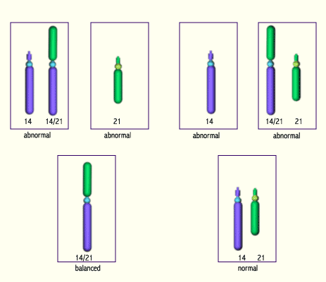
| Carriers of such robertsonian translocations are phenotypically inconspicuous. Also here, though, problems arise when it comes to gamete formation because, normally, the diploid chromosome set is halved thereby. Since, however, in this translocation a chromosome has fused to another one, no ordered segregation can take place. The carrier has a larger probability of having offspring with trisomy/monosomy and this is independent of his age. Often a translocation (e.g., t[14q21q]) is found in families with inherited trisomy 21. |
|
Fig. 16 - Robertsonian translocation
of acrocentric chromosomes |
|
Legend |
|
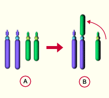
A
B
|
normales Chromosomenpaar
centric fusion of two
non-homologous chromosomes |
|
|
|
Fig. 16
In a reciprocal translocation two acrocentric chromosomes lose their short arms. Afterwards, the remaining partial pieces (q- arms) fuse to one another.
Animation (50 Kb)
|
| Fig. 17 - Gamete formation in acrocentric fusions |
|
Legend |
|
|
Fig. 17
This simplified model shows that in a robertsonian translocation the gamete formation is disturbed. On the one hand, monosomies or trisomies can arise and, on the other hand, gamete formation can also lead to normal or balanced gametes.
Animation (116 Kb)
|
|
Remarks
|
|
Various possibilities for the origin of a trisomy 21
- Non-disjunction of the chromosome 21 during meiosis = free form of the trisomy 21 = dependent of the maternal age (90%)
- Centric fusion of acrocentric chromosomes (robertsonian translocation) = inherited form of trisomy 21 = not dependent on maternal age (10%)
|
|
|
|
|
| In all structural aberrations the parental chromosome status should be examined. Carriers of balanced translocations are found very frequently in the population (1:500). Generally they are phenotypically inconspicuous and healthy, but they must be informed that their offspring can have unbalanced aberrations. A prenatal diagnosis is to be recommended. |
|
|
|
|
|
|