|

|
|
|
The follicle stages from primordial follicle to tertiary follicle
|
|
|
|
At the time of birth all the surviving primary oocytes are surrounded by thin, single layers of so-called follicular epithelial cells. These are delimited from the rest of the ovarian stroma by a thin basal lamina. Follicular epithelial cells are former coelomic epithelial cells. The primordial follicles always form the majority of the follicles in the ovary.
|
|
|
|
Under the influence of the sex hormones some of them are able to develop further to one or more of the subsequent stages in the following 50 years. Although this further development can already take place sporadically in the time before birth and up to puberty, the main part occurs as soon as a regular hormonal cycle is established. Particularly the last phase of the maturation of a tertiary follicle to become a large follicle, ready to rupture, remains reserved for the time of regular cycles. |
|
|
|
|
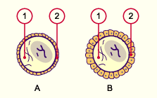
| In the transition of the primordial follicles into primary follicles the follicular epithelium that surrounds the oocyte becomes iso- to highly prismatic. |
|
Fig. 19 - Primary follicle |
|
Legend |
|
A
B
1
2
|
Primordial follicle
Primary follicle
Oocyte
Follicular epithelium |
|
|
|
Fig. 19
Scheme of the development from primordial follicle to primary follicle.
|
| When primary follicles survive, secondary follicles with follicular epitheliums encompassing multiple rows are engendered. This is now called the stratum granulosum. In the secondary follicles a glycoprotein layer, the pellucid zone, between the oocyte and follicular epithelium becomes visible. Cytoplasmic processes of the granulosa cells that lie upon it reach the oocyte through the pellucid zone and thereby assure their maintenance function. Outside the basal lamina the stroma ovarii organizes itself to become theca folliculi cells. |
|
Fig. 20 - Secondary follicle |
|
Legend |
|
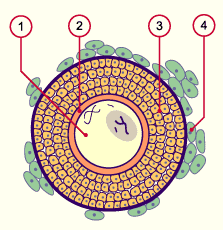
1
2
3
4
|
Oocyte
Pellucid zone
Stratum granulosum
Theca folliculi cells |
|
|
|
Fig. 20
Scheme of a secondary follicle:in the transition from primary to secondary follicle the stratum granulosum is engendered from the cells of the follicular epithelium. The stroma ovarii organizes itself around the secondary follicle to become the theca folliculi (interna and externa).
Histology of primordial, primary,and secondary follicles.
|
|
If the secondary follicles survive, tertiary follicles are engendered. Their identifying characteristic is a fluid-filled cavity, the antral follicle. The oocyte lies at the edge in a mound made of granulosa epithelial cells, the cumulus oophorus. In the meantime it has grown so large that its cellular nucleus has attained the size of a whole primordial follicle. The connective tissue around the follicle has already clearly differentiated itself into a theca interna, well supplied with capillaries, out of large, lipid-rich cells (hormone production) and a theca externa, which forms a transition to the stroma ovarii and contains larger vessels.
|
|
|
| Fig. 21 - Tertiary follicle |
|
Legend |
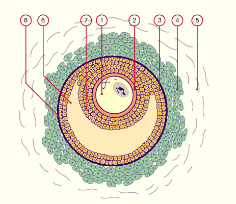
1
2
3
4
5
6
7
8
|
Oocyte
Pellucid zone
Stratum granulosum
Theca interna
Theca externa
Antral follicle
Cumulus oophorus (Granulosa cells, together with the oocyte)
Basal lamina between theca and stratum granulosum |
|
|
|
Fig. 21
In a tertiary follicle the theca can be subdivided into an interna (hormone production) and an externa (transition to the ovarian stroma).
Histology of tertiary follicles.
Histology of cumulus oophorus.
|
|
Decisive for a successful follicle growth is a well-developed net of capillaries in the theca interna. The precise steering mechanism that leads to the selection of a follicle and its subsequent maturation to become a graafian follicle is still unknown. Before ovulation a growth spurt of the tertiary follicles takes place.
|
|
|
|
This corresponds to an especially large tertiary follicle that can be expected to suffice for ovulation.
|
|
|
|
|