3.3 Spermatogenesis


Developmental stages of spermatogenesis

Quiz

Quiz 12


In the course of spermatogenesis the germ cells move towards the lumen as they mature. The following developmental stages are thereby passed through:

Quiz

Quiz 13


Quiz

Quiz 14


  • A-spermatogonium
  • B-spermatogonium
  • Primary spermatocyte (= spermatocyte order I)
  • Secondary spermatocyte (= spermatocyte order II)
  • Spermatid
  • Sperm cell (= spermatozoon)


The spermatogenesis can be subdivided into two successive sections:

  • The first comprises the cells from the spermatogonium up to and including the secondary spermatocyte and is termed spermatocytogenesis.
  • The second one comprises the differentiation/maturation of the sperm cell, starting with the spermatid phase and is termed spermiogenesis (or spermiohistogenesis).


The temporal course of spermatogenesis


The approximate 64 day cycle of the spermatogenesis can be subdivided into four phases that last differing lengths of time:


Mitosis of the spermatogonia 16 days Up to the primary spermatocytes
First meiosis 24 days For the division of the primary spermatocytes to form secondary spermatocytes
Second meiosis A few hours For engendering the spermatids
Spermiogenesis 24 days Up to the completed sperm cells
Total ~64 days    


Fig. 12 - The spermatogenesis generations  Legend

Fig. 12
The stem cell population of the germinal cells lies on the basal lamina of the convoluted seminiferous tubules.
These are Type A spermatogonia. These cells undergo mitosis: one of the daughter cells renew the stock of type A spermatogonia, the other becomes a type B spermatogonia.
These divide and their daughter cells migrate towards the lumen. In roughly 64 days they differentiate themselves thereby into sperm cells up to the outer surface of the epithelium (one should note that in these cellular divisions, the separation of the cytoplasm is not complete. Whole networks of connected cells arise. So, for example in the last generation, the spermatids, far more cells are bound to each other than as shown here).


Spermatocytogenesis


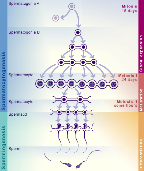
Among the spermatogonia (all in all, over 1 billion in both testicles) that form the basal layer of the germinal epithelium, several types can be distinguished: certain type A cells are seen as spermatogonia that divide mitotically and reproduce themselves (homonymous division), whereby the spermatogonia population is maintained.
The beginning of spermatogenesis is introduced through the so-called heteronymous division, in which the daughter cells (second group of type A cells) remain bound together by thin bridges of cytoplasm. Through the preservation of these cytoplasmic connections, spermatogonia are inducted into the spermatogenesis process.

After a further mitotic division type B spermatogonia are engendered that also divide themselves mitotically into primary spermatocytes (I).


The freshly created primary spermatocytes (I) now enter into the first meiosis. They then go immediately into the S phase (that is, into the preleptotene meiosis), double their internal DNA, leave the basal compartment and reach the special milieu of the luminal compartment. Following the S phase, these cells attain the complex stage of the prophase of the meiosis and become thereby noticeably visible with a light microscope.

This prophase, which lasts 24 days, can be divided into five sections:

  • Leptotene
  • Zygotene
  • Pachytene
  • Diplotene
  • Diakinesis

Commentary

In the heteronymous division the cytoplasmic division is not completed; the daughter cells stay bound together through thin cytoplasmic bridges.
Also in the subsequent meiosis the cytoplasmic division is incomplete, so that from one spermatogonium a network of daughter cells arises that doubles in size in each generation. The forming of such networks assures that all of the processes in each generation occur in step with each other.

In the prophase in every germ cell a new combination of maternal and paternal genetic material occurs. After the long prophase follow the metaphase, anaphase and telophase that take much less time. One primary spermatocyte yields two secondary spermatocytes.

Commentary

about meiosis


The secondary spermatocytes go directly into the second meiosis, out of which the spermatids emerge. Since in the secondary spermatocytes neither DNA reduplication nor a recombination of the genetic material occurs, the second meiosis can take place quickly. It lasts only around five hours and for that reason secondary spermatocytes are rather seldom seen in a histological section. Through the division of the chromatids of a secondary spermatocyte, two haploid spermatids arise that contain only half the original DNA content.

Besides the sperm cells the spermatids are the smallest cells of the germinal epithelium. In a process lasting several weeks (so-called spermiogenesis or spermiohistogenesis) they are transformed into sperm cells with the active assistance of the Sertoli's cells.


Previous page | Next page