| The paternal and the maternal pronuclei move towards each other with the help of microtubules, which begin to be formed immediately after impregnation, i.e., by the penetration of the spermatozoon. They grow in a star-like pattern out of the paternal centrosome directly beside the forming paternal pronucleus (= formation of an aster made of dozens of microtubules). The microtubular proteins themselves arise from the cytoplasma of the oocyte. |
|
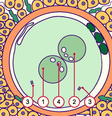
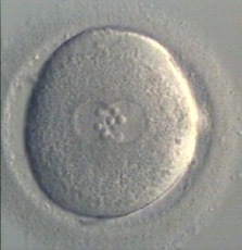
Fig. 46 - 6 hours after impregnation |
|
Legend |
|
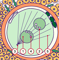
1
2
3
4
5
|
Paternal pronucleus
Maternal pronucleus
Paternal centrosome
"Inner bodies"
Maternal astral microtubule |
|
|
|
Fig. 46
Schematic diagram of an impregnated oocyte: An aster has formed out of what was originally the paternal centrosome (more info). In the pronuclei the so-called «inner bodies» can be seen, which are lined up as can typically be observed.
|