|

|
|
|
10.3 Placental blood circulation
|
|
|
Fetal and maternal blood circulation systems
|
|
|
|
The placental circulation brings into close relationship two circulation systems: the maternal and the fetal. The supply of blood to the placenta is influenced by various factors, especially by the arterial blood pressure, uterine contractions, tobacco abuse, medications and hormones. Placental blood flow is increased at term and amounts to 500 ml/min (80% of the uterine perfusion).
|
|
|
|
The fetal circulation system
|
|
|
The villus capillaries are branches of the umbilical vessels. Fetal blood comes via the two Aa .umbilicales in the villi and leaves the placenta through a single navel vein, the vena umbilicalis. Their supply amounts to approximately 40% of the fetal heart blood volume per minute.
The blood pressure in the arteria umbilicalis amounts to 50 mmHg and the blood flows through finer vessels that cross through the chorionic plate to the capillaries in the villi where the arterial blood pressure falls to 30 mmHg. In the umbilical vein the pressure is 20 mm Hg. The pressure in the fetal vessels and their villus branches always lies over that of the intervillous space. This protects the fetal vessels from collapse (interactive diagram).
|
|
|
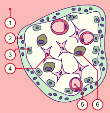
| Fig. 33 - Utero-placental circulation system |
|
Legend |
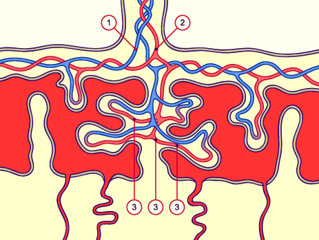
1
2
3
|
Umbilical arteries
Umbilical vein
Fetal capillaries |
|
|
|
Fig. 33
Fetal blood flows via the Aa. umbilicales, where the blood pressure amounts to 50 mm Hg, through finer vessels in the chorionic plate, in order to finally reach the capillaries in the villi, where the pressure sinks to 30 mm Hg.
Note: Due to their low oxygen content, the fetal Aa. umbilicales are shown in blue and the oxygen-rich veins in red.
|
|
The maternal circulation system
|
|
|
During the pregnancy the uterine circulation constantly adapts in order to be adequate for the growing metabolic needs of the embryo. Via the spiral arteries (80 -100 mm Hg) that come from the uterine arteries (Aa. uterinae), maternal blood gets into the intervillous spaces in a region delimited by the anchoring villi. Subsequently the blood leaves the intervillous spaces via the uterine veins that are arranged in the periphery of the intervillous space.
The flow of the placental blood amounts to 600 cm3/min and the pressure in the spiral arteries to 70 mm Hg. In the intervillous spaces the pressure falls to only 10 mm Hg .The blood in the intervillous space is exchanged 2-3 times per minute.
|
|
|
| Fig. 34 - Maternal circulation system |
|
Legend |
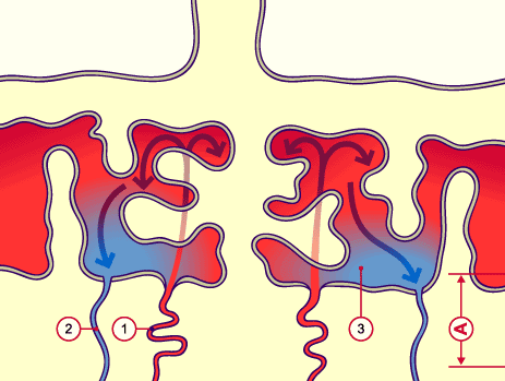
1
2
3
A |
Spiral arteries
Uterine veins
Intervillous spaces
Basal plate |
|
|
|
Fig. 34
Maternal blood arrives at the intervillous space via arteries that open directly into the intervillous space. At the placental level, it thus finds itself temporarily outside the vessel network.
|
|
The placental barrier is composed of structures that separate the maternal and the fetal blood .The makeup of the placental barrier changes over the course of the pregnancy.
|
|
|
| In the first trimester it consists of the syncytiotrophoblast, the cytotrophoblast (Langhans' cells), the villus mesenchyma (in which numerous ovoid Hofbauer cells that exhibit macrophage properties are found) and the fetal capillary walls. |
|
Fig. 35 - Villus in the first trimenon |
|
Legend |
|
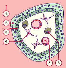
1
2
3
4
5
6
|
Intervillous space
Syncytiotrophoblast
Cytotrophoblast
Villus mesenchyma
Fetal capillaries
Hofbauer macrophages |
|
|
|
Fig. 35
The villus has an intact syncytio-
and cytotrophoblast layer. In the villus interior there are mesenchymal cells with macrophages and fetal capillaries.
|
|
|
|
| During the 4th month the cytotrophoblast disappears from the villus wall (interactive diagram) and the thickness of the barrier decreases while the surface area increases (roughly 12 m2 towards the end of the pregnancy). In the 5th month the fetal vessels have multiplied their branches and gotten closer to the villus surface. |
|
Fig. 36 - Villus in the second trimenon |
|
Legend |
|
1
2
3
4
5
6 |
Intervillous space
Syncytiotrophoblast
Cytotrophoblast
Villus mesenchyma
Fetal capillaries
Hofbauer macrophages |
|
|
|
Fig. 36
In the middle third of the pregnancy the capillaries migrate to the villus surface. The cytotrophoblast layer disappears slowly and the syncytiotrophoblast layer becomes thinner.
|
|
|
|
|
During the 6th month the nuclei of the syncytiotrophoblast group together in the so-called proliferation knots. The other zones of the syncythiothrophoblast lack nuclei and are adjacent to the capillaries (exchange zones).
|
|
|
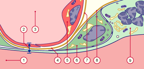
| Fig. 37 - Placental barrier in a mature villus |
|
Legend |
1
2
3
4
5
6
7
8
9 |
Intervillous space (with maternal blood)
Placental barrier of a terminal villus
Fetal capillaries
Merged basal membranes of the fetal capillary and of the syncythiothrophoblast
Endothelial cells
Rare cytotrophoblast cells
Basal membrane of the capillaries
Basal membrane of the trophoblast portion
Syncytiotrophoblast with proliferation knots (nuclei rich region) |
|
|
|
Fig. 37
At its thinnest part, the placental barrier is reduced to the nucleus-free syncytiotrophoblast, the merged basal membrane and the endothelium.
|
|
|