|

|
|
|
Development of the umbilical cord
|
|
|
|
The umbilical cord is formed when the body stalk and the ductus omphalo-entericus as well as the umbilical coelom are enveloped by the spreading amnion between the 4th and 8th week. Finally, when the membranes of the amniotic cavity come into contact with those of the chorionic cavity and the two extra-embryonic mesoderm layers that cover both membranes, fuse. With the flexing movements of the embryo, the amnion encircles the body stalk, the ductus omphalo-entericus and the umbilical vessels, thus circumscribing the elements of the umbilical cord.
|
|
|
Animation
|
|
Development of the embryo from the trilaminar embryonic disk up to the end of the embryonic period
(the same animation is involved, but with varying quality.)
706 Kb, 1.3 Mb
|
|
|
| Fig. 48 - Body stalk |
|
Fig. 49 - Formation of the umbilical cord |
|
Legend |
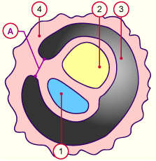
A
B
|
Body stalk
Stem of umbilical vesicle |
|
|
|
1
2
3
4
5
|
Amniotic cavity
Umbilical vesicle
Chorionic cavity
Villous chorion
Allantois |
|
|
|
Fig. 48
Body stalk at around the 3rd week
Fig. 49
Formation of the umbilical cord at around the 3.5th week
|
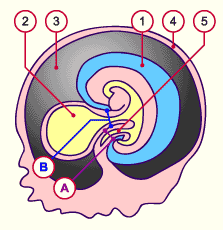
| Fig. 50 - Umbilical cord |
|
Fig. 51 - Umbilical vesicle in the chorionic cavity |
|
Legend |
A
B
C
|
Body stalk
Stem of umbilical vesicle
Umbilical cord |
|
|
|
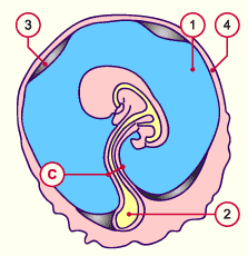
1
2
3
4
|
Amniotic cavity
Umbilical vesicle
Chorionic cavity
Chorion laeve |
|
|
|
Fig. 50
The body stalk and the yolk stalk are now united and form the umbilical cord. Through increasing secretion of amniotic fluid the chorionic cavity becomes obliterated. Here at around the 4.5th week: The chorionic cavity is reduced in size
Fig. 51
Flexing of the embryo at around the 8th week with expansion of the amnion that encircles the body stalk and the ductus
omphalo-entericus, the umbilical coelom and the umbilical vessels
|
|
In the early stage (at around the 8th week) the umbilical cord is in the form of a very thick and short section with the following structures:
- The ductus omphalo-entericus which connects the primitive intestines with the umbilical vesicle and two vitelline vessels (vasa omphalomesenterica, 2 arteries and 2 veins!). The umbilical vesicle is located in the chorionic cavity (exocoelom = extra-embryonic coelom).
- The body stalk with the allantois, the umbilical vessels (2 arteries and 1 vein!). During the development it gets shifted ventrally in order to finally fuse with the stem of the umbilical vesicle.
- The umbilical coelom that connects the extra-embryonic coelom with the intra-embryonic coelom
|
|
|
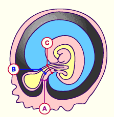
| Fig. 52a - Development |
|
Fig. 52b - Development |
|
Legend |
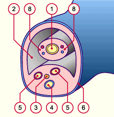
1
3
4
5
6 |
Ductus omphalo-entericus
Allantois
Umbilical vein
Umbilical arteries
Amnion |
|
|
|
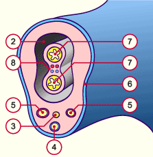
2
7
8
|
Extra-embryonic coelom
(umbilcal coelom)
Intestinal tube
(physiologic umbilical hernia)
Vasa omphalomesenterica |
|
|
|
Fig. 52a
Transversal section of the primitive umbilical cord after approximately 8 weeks
Fig. 52b
Transversal section of the primitive umbilical cord with physiologic umbilical hernia at around the 3rd month
|
|
Further development promotes both lengthening as well as the reduction of some structures.
|
|
|
|
Lengthening:
The amniotic cavity forms a covering around the ductus omphalo-entericus and the body stalk, which elongates. The newly formed umbilical cord continues to lengthen to allow for fetal movements and coils in the amniotic cavity.
|
|
|
|
Reduction:
Numerous elements degenerate in the 3rd month.
- the omphalo-enteric duct (it can remain in the form of a Meckel's diverticulum)
- the umbilical vesicle of the allantois (it is obliterated in order to form the umbilical ligament, lying medially in adults)
- the vitelline circulation system in the extra-embryonic region.
- the umbilical coelom, which clumps and disappears.
Finally, only the body stalk remains with its umbilical vessels (2 arteries, 1 vein), which are surrounded by an amniotic epithelial layer. The connective tissue of the body stalk and the amnion (that stems from the extra-embryonic mesoblast) go over into a common umbilical cord connective tissue, the so-called "Wharton jelly", an elastic and resistant tissue that protects the umbilical vessels from possible mechanical pressure and creasing.
|
|
|
|
|