|

|
|
|
6.1 Role and functional anatomy of the endometrium
|
|
|
Cyclic hormonal alterations of the endometrium
|
|
|
|
Over the whole sexually active time span (from puberty to the menopause) the endometrium is subject to cyclic changes under the influence of the same hormones that regulate ovulation. There are three levels of this hormonal regulation: hypothalamus-pituitary-ovary and it takes place via longer and shorter feedback mechanisms.
|
|
|
|
The menstruation phase (1rst to the 4th day) distinguishes the beginning of each menstruation cycle. When an implantation does not occur, the back-formation of the yellow body (corpus luteum) lowers the amounts of circulating estradiol and progesterone hormones, which leads to the expulsion of the functional layer of the endometrium.
|
|
|
Fig. 4 - Endometrium in the
menstruation phase |
|
Fig. 5 - Enlargement |
|
Legend |
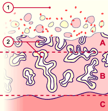
A
B
C
1
2
|
Functional layer
Basal layer
Myometrium
Uterine cavity with epithelial cells,
blood corpuscles and remainders
of the expulsed mucosa
Intact and partially expulsed uterine glands |
|
|
|
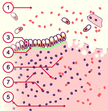
3
4
5
6
7
|
Intact epithelial cells
Basal membrane
Uterine stroma
Blood corpuscles
Free cells of the connective tissue |
|
|
|
Fig. 4, Fig. 5
Expulsion of the functional layer of the endometrium (spongiosa and compacta) mixed with blood, endometrial debris and lymphocytes.
|
|
More info
|
|
Vascular mechanisms basic to menstruation
The mechanisms that regulate the menstruation phase (1rst-4th day) result from the reduction in the estrogen and progesterone values, leading to a constriction of spiral arteries and consequent necrosis of the tissue.
Only the functional layer of the endometrium is affected by these cyclic changes - the basal layer remains intact.
The uterine vessel network (scheme) exhibits a selective sensibility with regard to the cyclic hormonal alterations. The radial and basal arterioles do not react to the hormonal variations, whereas the spiral arteries of the functional layer are hormone sensitive and constrict when the progesterone concentration decreases. 2
The cramp-like contractions of the tunica media of the spiral arteries is responsible for an interruption of the blood supply (ischemia), which results in the dying out of the functional layer of the endometrium. Together with blood, which does not coagulate due to a local fibrinolytic factor, the necrotic tissue is eliminated (menstruation).
|
|
|
|
|
|
The follicular or proliferative phase
|
|
|
During the proliferative or follicular phase (4th to 14th day) the secretion of estrogen through the growing ovarian follicle is responsible for the proliferation of the endometrium (intensive mitosis in the glandular epithelium and in the stroma).
The uterus epithelium clothes the surface again. In this stage a certain number of epithelial cells equipped with cilia can be recognized.
The glands grow longer and the spiral arteries wind themselves lightly into the stroma. At the end of the proliferative phase the estradiol peak (released by the growing follicles) triggers a positive feedback mechanism at the level of the pituitary and the ovulation commences 35 to 44 hours after the initial LH increase (cyclic hormonal changes).
|
|
|
Fig. 6 - Endometrium in the early
proliferative phase
|
|
Fig. 7 - Uterine glands
|
|
Legend |
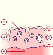
1
2
3
4 |
Glandular epithelium
Endometrium that is a little
developed
Uterine glands
Myometrium |
|
|
|
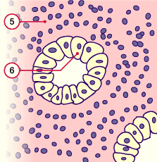
5
6
|
Stroma of the endometrium
Epithelial cells of the uterine glands |
|
|
|
Fig. 6, Fig. 7
Early proliferative phase,
characterized by a thin, relatively uniform endometrium.
|
More info
|
|
Histology in slight and increased enlargement
|
|
|
Fig. 8 - Endometrium in the late
proliferative phase
|
|
Fig. 9 - Uterine glands
|
|
Legend |
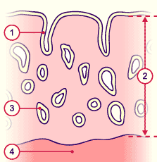
1
2
3
4
|
Glandular epithelium
Endometrium during the
proliferation
Uterine glands
Myometrium |
|
|
|
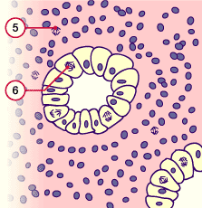
5
6
|
Stroma of the endometrium
(mitosis)
Epithelial uterine gland cells with mitotic figures |
|
|
|
Fig. 8, Fig. 9
Late proliferative phase:
thickened endometrium with an increased number of glands and mitosis, visible in the glandular epithelium and stroma.
|
|
The luteinizing or secretory phase
|
|
|
During the secretory or luteinizing phase (14th to 28th day) the endometrium differentiates itself due to the influence of progesterone (from the corpus luteum) and attains its full maturity. The glands and arteries begin to entwine. The connective tissue stroma becomes the place of edematous changes.
The time period of the maximal reception ability for the blastocyst lies between the 20th and the 23rd day. This phase of the endometrium lasts 4 days and is usually termed the "implantation window" .
|
|
|
Fig. 10 - Endometrium in the early secretory phase
|
|
Fig. 11 - Uterine glands
|
|
Legend |
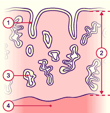
1
2
3
4
|
Glandular epithelium
Thickened endometrium
Uterine glands, curled
Myometrium |
|
|
|
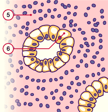
5
6
|
Stroma of the endometrium
Epithelial uterine gland cells with
glycogen collections at the basal
pole |
|
|
|
Fig. 10, Fig. 11
Early secretory phase:
the endometrium nears its full maturity. The nuclei of the epithelial cells are round and, due to the important production and storage of glycogen at the basal pole, lie at the apical pole near the lumen.
|
More info
|
|
Histology
slight and increased enlargement
|
|
|
Fig. 12 - Endometrium in the middle
secretory phase
|
|
Fig. 13 - Uterine glands
|
|
Legend |
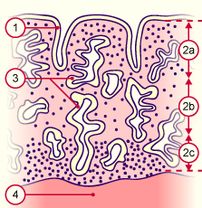
1
2a
2b
2c
3
4
NB |
Glandular epithelium
Stratum compactum
Stratum spongiosum
Stratum basale
Curled uterine glands
Myometrium
2a + 2b = Stratum functionale
|
|
|
|
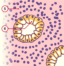
5
6
|
Stroma of the endometrium
Epithelial cells of the uterine glands
with glycogen collections at the
apical pole |
|
|
|
Fig. 12, Fig. 13
Middle secretory phase:
The endometrium is now mature; the glycogen migrates from the basal to the apical pole, whereby the nuclei of the epithelial cells are shifted to the basal pole.
The secretion containing glycogen is released into the glandular lumen.
|
|
|

