|

|
|
|
|
Hemoglobin in human erythrocytes consists of 2 paired globin chains that are arranged around a heme that, for its part, can bind oxygen. While only hemoglobin of the Hb Gower 1 (z2e2) type with z and e chains is formed in the blood islands of the umbilical vesicle, the mature erythrocyte of the hepatic phase exhibits mainly HbF (a2g2), the globin of which possesses a and g chains and represents the most important fetal hemoglobin.
|
|
|
|
More info
|
|
The transition from Hb Gower 1 to HbF takes place in steps in that both hemoglobins with a2e2 chains --> Hb Gower 2 as well as such with z2g2 chains --> Hb Portland can be found in the intermediate phase. The genetic information for the synthesis of the individual globin chains is found on chromosomes 11 and 16 (interactive diagram, 29.6 kb).
|
|
|
|
|
| Following birth, the fetal HbF is slowly replaced by the adult HbA1 (a2b2). This consists of a and b chains and represents the most important adult hemoglobin. In addition there is still a small portion of HbA2(a2d2) that exhibits d chains instead of b ones. |
|
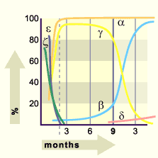
Fig. 4 - The formation of various
Hb-chains during the pregnancy |
|
Legend |
|
|
|
Fig. 4
Synthesis of the globin chains over the course of the pregnancy. In the embryonic period the e and z globin chains dominate. They are, though, rapidly taken over by a and g globin chains. Towards the end of the pregnancy, the g chains of the fetal hemoglobin are slowly replaced by the b chains (adult hemoglobin).
|
Physiology of the prenatal oxygen supply
|
|
|
|
The prenatal oxygen supply gets optimized by three factors:
|
|
|
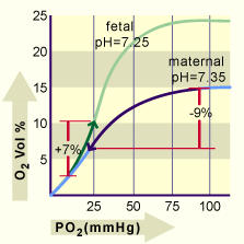
| All three factors are important for an efficient gas exchange and play a role in the different compartments of the maternal and infant blood. In the following diagram the O2 binding curves of the maternal and fetal blood at the time of birth are shown for the mean pH value in the placenta. In order to take into account the various hemoglobin concentrations of mother and fetus (12g/% and 18g/%, respectively), not the O2 saturation is displayed on the ordinate but rather the O2 concentration. |
|
Fig. 5 - O2 concentrations as function
of the O2 partial pressure |
|
Legend |
|
|
|
Fig. 5
The O2 concentrations as a function of the O2 partial pressure (PO2) for the maternal and fetal blood at the time of birth are shown on the two lines. During the gas exchange process in the placenta the O2 concentration in the maternal blood sinks around 9 volume %, while that of the fetal blood increases around 7 vol.%.
Please note that, for the same partial pressure (e.g. 50 mm Hg), fetal blood can bind considerably more O2 (22%) than that of the mother (12%).
|
|
|