|

|
|
|
|
The human body possesses two large cavities (pleural and peritoneal) and a smaller cavity (pericardial), all of which are covered with a mesothelium. One speaks of visceral mesothelium that coats the organs and parietal mesothelium that coats the somatic walls delimiting the cavitiy towards the inside and also the outside.
|
|
|
Origin of the pleural cavities
|
|
|
|
In stage 10 (28 days)  10 10 the cranial,section of the intraembryonic coelom consists of a middle part, the pericardial cavity, and two thin canals laterally, the pericardioperitoneal canals. the cranial,section of the intraembryonic coelom consists of a middle part, the pericardial cavity, and two thin canals laterally, the pericardioperitoneal canals.
|
|
|
|
They connect the pericardial cavity with the part of the intraembryonic coelom that is open towards the outside, the future peritoneal cavity. At this point no pleural cavity yet exists because the lungs have not yet begun to develop.
|
|
|
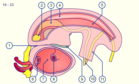
| Fig. 14 - Intraembryonic coelom in stage 12 |
|
Legend |
1
2
3
4
5 |
Pharyngeal pouch
Aortic arch
Pericardioperitoneal canal
Peritoneal cavity
Aortic sac |
|
|
6
7
8
9 |
Pericardial cavity (without heart)
Venous sinus
Liver bud
Umbilical coelom |
|
|
|
|
|
Fig. 14
In stage 12 the peritoneal cavity is connected with the pericardial cavity via the pericardio-
peritoneal canals.
From the foregut in the middle, the lung buds will soon sprout on both sides into the pericardio-
peritoneal canals. Observe the proximity of the heart and liver anlagen.
|
|
|
|
|
In stage 13 (32 days) 13 the lung buds grow into the pericardioperitoneal canals and dent them. Thereby the pericardioperitoneal canals are subdivided on both sides by these lung buds that are sprouting in from the medial direction
|
|
|
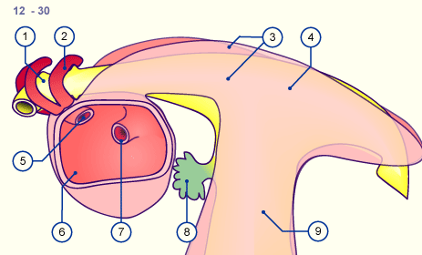
| Fig. 15 - Schematic cross-section through an embryo in stage 14 |
|
Legend |
1
2
3
4
5
6
7
8
9
10 |
Arm buds
Pleuropericardial membrane
Development of the pleural cavity in the pericardioperitoneal canal
Esophago-tracheal septum
Esophagus
Trachea
Lung buds
Bulbus cordis
Heart
Pericardial cavity |
|
|
|
Fig. 15
The cross-section of this embryo in stage 14 shows the lung buds that grow into the pericardio-
peritoneal canal on both sides. |
|
With the increase in size of the lung anlage the pericardioperitoneal canal widens to become the pleural cavity that is separated from the pericardial cavity by the pleuropericardial membrane and from the peritoneal cavity by the pleuroperitoneal membrane.
|
|
|
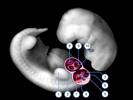
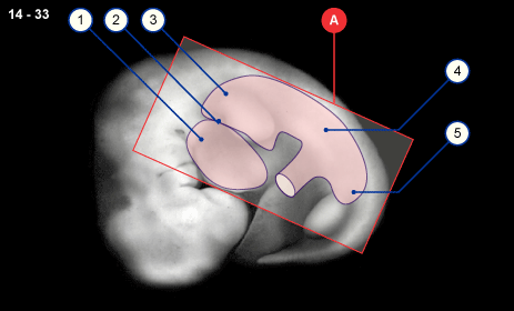
| Fig. 16 - Lateral section of an embryo in stage 14 |
|
Legend |
1
2
3
4
5
|
Pericardial cavity
Pleuro-pericardial canal / pleuropericardial membrane
Pleural cavity
Peritoneal cavity
Pelvic part of the peritoneal cavity |
|
|
|
Fig. 16
Intraembryonic coelom projected on an embryo in stage 14. |
|
In the region of the peritoneal cavity and the umbilical coelom, the left and right peritoneal tubes join ventrally to form a common cavity, the peritoneal cavity, that goes over into the umbilical coelom. In the dorsal region they form the meso of the guts.
|
|
|
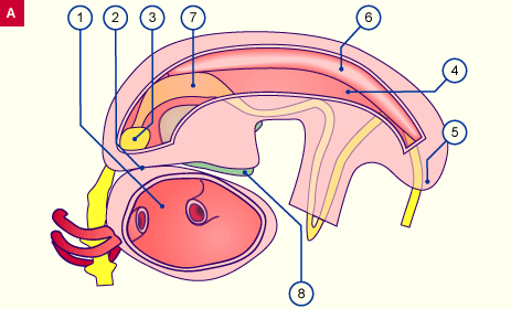
| Fig. 17 - Cut-open cavities with view the with visceral serosa-coated organs |
|
Legend |
1
2
3
4
5
6
7
8 |
Pericardial cavity
Pleuro-pericardiac canal / pleuro-pericardial membrane
Lungenanlage
Peritoneal cavity
Pelvic part of the peritoneal cavity
Urogenital ridge
Stomach
Liver |
|
|
|
Fig. 17
Enlarged, more detailed view of insert A from Fig. 16.
In the cut-open coelom cavity one sees the intraperitoneal organs that are coated with mesothelium: the stomach and liver, as well as the urogenital ridge. |
|
The lungs become covered by the visceral layer of the pleural cavity, the pleura visceralis. Towards the outside, the pleural cavity is bounded by the parietal layer, the pleura parietalis. Through their rapid increase in size the two lungs, left and right, enclose the heart that is in their middle.
In the following interactive diagram (development of the pleural cavity in various stages) the intraembryonic coelom can be seen in various stages. One distinguishes the pericardial cavity in the middle from the two pleural cavities and, in the intestinal region, the peritoneal cavity
|
|
|
|
|