|

|
|
|
4.2 Getting the spermatozoa ready
|
|
|
Processes in the male genital tract
|
|
|
|
The supplying of the spermatozoa occurs in the male genital tract. Figure 21 provides an overview of the path the spermatozoa travel during an ejaculation.
|
|
|
The spermatozoa are produced from the spermatogonia of the testis. They arise through mitotic and meiotic divisions in the seminiferous tubules (tubuli seminiferi contorti ) there (see spermatogenesis).
Around 600 - 800 seminiferous tubules build the main part of the testis parenchyma and are arranged in over 300 tiny, incompletely partitioned lobules. With a length of roughly 50 - 60 cm for an individual seminiferous tubule it can be calculated that in a single testis a total canal length of around 300 m is accommodated. In addition, the cells that produce the hormone testosterone (Leydig's cells) are present in the interstitial tissue as well. |
|
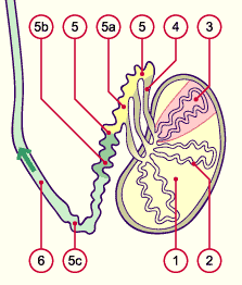
Fig. 22 - Testicle and epididymis |
|
Legend |
|

1
2
3
4
5
5a
5b
5c
6
|
Testis
Seminiferous tubules
(tubuli seminiferi contorti)
Lobules
Ductuli efferentes testis
Ductus epididymidis
Caput epididymidis
Corpus epididymidis
Cauda epididymidis
Deferent duct / vas deferens |
|
|
|
Fig. 22
In the testicle the tightly coiled seminiferous tubules (tubuli seminiferi contorti) are arranged in lobules.
The epididymis is formed from one, single 5-6 m long, tightly coiled canal, termed ductus epididymidis. The epididymis is subdivided into a head (caput) - in this part the 10-20 ductuli efferentes testis coming from the testis flow - a body (corpus) and a tail (cauda).
|
|
Processes in the epididymis
|
|
|
Following their production in the seminiferous tubules of the testis, the sperm cells are collected and stored in the ductus epididymidis.
|
|
|
Maturation steps of the spermatozoa in the epididymis:
- Through the deposition of new proteins in the nucleus, the DNA becomes more condensed. The sperm head becomes smaller, thereby, and more compact. This is an important step for the later correct decondensation of the paternal DNA in the maternal oocyte.
- The already meager cytoplasma is further reduced, making the sperm cells more slender.
- The ability for motility is achieved but at the same time inhibited by the milieu.
- The structure of the plasma membrane is altered. This has effects on the motility, the capacitation ability and the ability for the acrosome reaction.
|
|
|
|
|
|
From this, the following should be noted:
|
|
|
| Only spermatozoa that have passed through the epididymis are mature enough to be capable of motility. |
|
|
|
|
|
|

