|

|
|
|
|
The fertilization process takes place in the female genital tract.
|
|
|
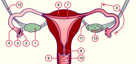
| Fig. 5 - The female genital tract |
|
Legend |
1
2
3
4
5
6
7
8
9
10
11
12
13
|
Ovary
Infundibulum
Fimbriae
Fallopian or uterine tube
Ampullary part of the tube
Uterine musculature
Uterine mucosa
Cervix
Portio
Vagina
Ligamentum ovarii proprium
Suspensory ligament of the ovary
Ovary cut open (follicles in various stages) |
|
|
|
Fig. 5
Schematic drawing of the female sexual organs. Frontal view: vagina, uterus; right tube and right ovary are cut open.
|
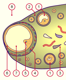
The ovary and the dominant follicle
|
|
|
| Roughly a week before the midpoint of the menstrual cycle the dominant follicle develops in one of the two ovaries. This grows faster than the other tertiary follicles and prepares itself for ovulation. It reaches a diameter of up to 25 mm and is also known then as the graafian follicle. |
|
Fig. 6 - Graafscher Follikel |
|
Legend |
|
1
2
3
4
5
6
7
8 |
Tertiary follicle
Ovary cortex
Medulla of the ovary
Theca with capillary network of
the graafian follicle
Oocyte within the cumulus
oophorus
Stratum granulosum
(granulosa cells)
Antrum folliculi with follicle fluid
Peritoneal cavity |
|
|
|
Fig. 6
Schematic diagram of a graafian follicle in the ovary.
|
The ultrasound image in Fig. 7 shows the follicle fluid as a black surface. The recording was made shortly before ovulation. Since at this moment the cumulus oophorus has detached itself from the granulosa, the oocyte "swims" surrounded by its cumulus cells (so-called corona radiata) in the follicle fluid. This cannot be seen, though, in the figure.
The graafian follicle bulges out the ovary's surface. It is ready to tear open, i.e., its wall and the peritoneal covering will soon rupture and its watery contents will pour into the infundibulum of the tuba, together with the oocyte. The diameter of the follicle that is about to rupture is here 22 mm (corresponding to 5 ml of follicle fluid). |
|
Fig. 7 - Ultrasound image of a
graafian follicle |
|
Legend |
|
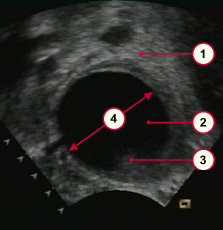
1
2
3
4
|
Ovarian tissue
Follicle about to rupture
Remains of the cumulus oophorus
The diameter of the follicle is of 22 mm |
|
|
|
Fig. 7
Single frame from a series of video images that shows the follicle about to rupture
© Dr. L. Rayo, Inselspital Bern
Video (150 kB)
|
|
A hormonal regulation is responsible for follicle maturation. The selection criteria for choosing the dominant follicle are still unknown, however. The following plot shows the course of the concentrations of the hormones that are involved during a monthly cycle.
|
|
|
| Fig. 8 - Hormone concentrations during a cycle |
|
Legend |
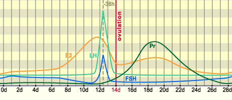
LH
E2
Pr
FSH
|
Luteinisierendes Hormon
Östradiol
Progesteron
Follikel stimulierendes Hormon |
|
|
|
Fig. 8
The curves show the concentrations of the FSH, LH, estradiol, and progesterone hormones in the blood. They have been recorded for the average period duration of 28 days. Roughly 38 hours before ovulation, a LH peak occurs.
|
|
Approximately one-and-a-half days before the midpoint of the cycle, the concentration of the luteinizing hormone (LH) rises steeply. The LH peak is responsible for a whole series of processes: triggering ovulation, creating an optimal milieu for a successful fertilization, and harmonizing the temporal sequence of the processes with one another.
With the LH peak the following maturation steps are now triggered in and around the oocyte - up to ovulation:
|
|
|
|
|
Commentary
|
|
What causes the LH peak?
What are the hormonal relationships that lead to the LH peak? How do they influence the follicle in its maturation process?
More about this in an interactive popup. (620 kB!)
|
|
|
|
More detail about these effects is provided in the next pages.
|
|
|
|
|