|

|
|
|
Ovulation, i.e., the emergence of the secondary oocyte from the follicle, depends on the disintegration of the follicle wall and the rupture of the ovarian surface.
A few hours after the FSH/LH peak one observes increased vascularization and edematous changes in the dominant follicle's surroundings. With time it is displaced towards the ovarian surface where it finally bulges out. |
|
Fig. 15 - Graafian follicle |
|
Legend |
|
1
2
3
4
5
|
Fallopian tube
Fimbriae
Ovary
Follicle
Stigma
|
|
|
|
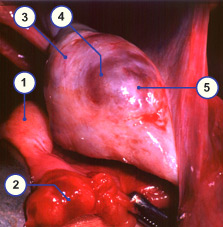
Fig. 15
Graafian follicle bulge on the ovary surface. The beginning formation of a stigma (whitish region free of blood vessels).
|
Video
|
|
Laparoscopic
video-recording of an ovary with follicle that is about to rupture (190 kB)
© PD. Dr. med. Michel Müller, Frauenklinik Inselspital Bern
|
|
|
|
The hyaluronic acid, secreted by the granulosa cells, has the property that it binds water molecules: The more hyaluronic acid is made, the more water can be absorbed. In this way, a rapid increase of the amount of follicle fluid occurs and this leads to a dramatic increase of the tension in the follicle wall. This together with the effects of a lytic enzyme finally leads to the rupture of the follicle at a narrowly circumscribed place.
On the ovarian surface above the follicle that is about to burst, a white point forms shortly before the rupture (due to compression of the blood vessels), the so-called stigma.
|
|
|
The acquisition of the oocyte by the fallopian tube
|
|
|
|
In the meantime, the fimbriae of the infundibulum have placed themselves around the ovarian stigma and have sealed this location off.
Via a rotation about the axis of the suspensory ligament of the ovary and the ovarian ligament the ovary can turn the follicle that is about to rupture towards the fallopian tube.
|
|
|
| Fig. 16 - Oocyte acquisition mechanism |
|
Fig. 17 - Oocyte acquisition mechanism |
|
Legend |
1
2
3
4
|
Ovary
Follicle that is about to rupture
Infundibulum
Fimbriae |
|
|
|
5
6
7
|
Fallopian tube
Ligamentum ovarium proprium
Suspensory ligament of the ovary |
|
|
|
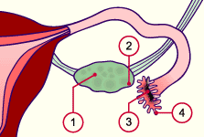
Fig. 16
Mechanism of oocyte acquisition:
Initial positions
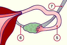
Fig. 17
Mechanism of oocyte acquisition:
Fimbriae apposed
|
|
When the surface ruptures the mass of cumulus cells, which are saturated with hyaluronic acid and which shelter the oocyte, reach the fallopian tube together with serous yellow follicle contents.
|
|
|
Video
|
|
Laparoscopic recording of the uterus, the fallopian tubes and the ovaries (Video: 620 kB)
© PD. Dr. med. Michel Müller, Frauenklinik Inselspital Bern
|
|
|
| Fig. 18 - Oocyte acquisition mechanism |
|
Legend |
1
2
3
4
5
6
7
8
9
10 |
Fallopian tube cut open with the tube mucosa that lies in folds
Closely apposed fimbriae
Follicle fluid that has flowed out
Secondary oocyte with corona radiata
Ovary with follicles in various stages of development and atresia
Pellucid zone
First polar body
Secondary oocyte
Cells of the corona radiata
Arrested spindle apparatus |
|
|
|
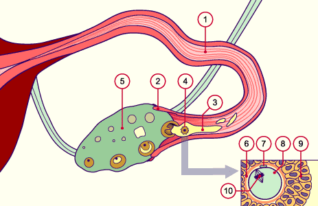
Fig. 18
Mechanism of oocyte acquisition:
Fimbriae and cutaway fallopian tube with oocyte and follicle fluid in it.
|
|
The outflow of the follicle contents is a procedure accompanied by coagulation processes. The protracted expulsion of the oocyte from the follicle makes it understandable that the fimbria funnel of the fallopian tube has enough time to flare itself over the ovulation location and to take up the oocyte-cumulus-complex with the accompanying volume of follicle fluid in the ampulla of the tube.
|
|
|
The oocyte in the cloud of cumulus cells following ovulation
|
|
|
| In the fallopian tube, the secondary oocyte is surrounded by the corona radiata and scattered parts of cumulus cells (so-called cumulus cell cloud). The fluid that lies in between is sticky and stringy (effect of the hyaluronic acid) with a high concentration of progesterone (to attract the spermatozoa). |
|
Fig. 19 - Oocyte following ovulation |
|
Legend |
|
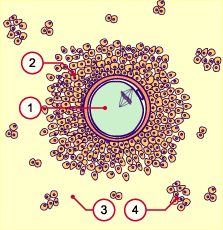
1
2
3
4
|
Secondary oocyte (in arrested
metaphase of the 2nd meiosis)
Corona radiata
Follicle fluid
Scattered groups of cumulus cells |
|
|
|
Fig. 19
Schematic of the oocyte following ovulation.
|
Video
|
|
The cloud of cumulus cells, the oocyte with the corona radiata, and the effect of the hyaluronic acid are depicted in vivo in this video (332 kB).
Recorded with the kind support of Dr. A. Senn, Lausanne und S. von Wyl, Bern
|
|
|
|
Commentary
|
|
The oocyte now "waits" in the fallopian tube on fertilization by the sperm. The matrix of hyaluronic acid holds it "captive" there, so to speak. After a number of hours the matrix liquefies more and more and the oocyte is gradually transported towards the uterus by the ciliary beats of the tube's epithelium cells. Since after ovulation the oocyte can only be fertilized within a few hours, the fertilization must almost inevitably take place in the ampullary part of the fallopian tube.
|
|
|
|
|
|
|