|

|
|
|
5.1 The cleavage divisions and the migration of the embryo through the tube
|
|
|
The polarity of the embryo
|
|
|
| The polarity of the embryo is seen in the forming of embryonic and abembryonalic poles. This is obvious when observing a blastocyst where an inner cell mass (ICM) has formed. This is concentrated at one pole in the interior of the hollow sphere and is made from blastomeres. |
|
Fig. 14 - Blastocyst on the 5th day |
|
Legend |
|

1
2
|
Blastomeres of the trophoblast
(abembryonic pole)
Inner cell mass
(ICM = embryoblast)
(embryonic pole) |
|
|
|
Fig. 14
Blastocyst that developed from an artificially fertilized oocyte, shortly before being introduced into the patient.
© Dr. S.v.Wyl, Frauenklinik Bern
|
| This polarity was already set up in the fertilized oocyte. Its cytoskeleton is substantially involved with this phenomenon since the cytoskeleton is anchored at the oolemma. If the observation conditions are favorable, it is possible to distinguish between a somewhat smoother and a somewhat rougher surface on the fertilized oocyte. These two differing surfaces reflect the polarity of the future embryo. |
|
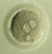
Fig. 15 - Impregnated oocyte |
|
Legend |
|
|
|
Fig. 15
In this picture, focused on the oocyte surface, one sees that the right hemisphere is smoother, while the left one exhibits a rougher surface structure. In the depth of the picture, two pronuclei in the typical vis-à-vis position and a polar body can be discerned.
|
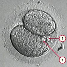
| The division of the zygote into the two-cell stage happens precisely so that one of the cells being formed is oriented to one pole and the other to the other one. The division always takes place so the polar bodies end up lying in the cleavage crease. This is not surprising since the cytoskeleton and the mitotic spindle function together to expel the polar bodies. |
|
Fig. 16 - Two-cell embryo |
|
Legend |
|
|
|
Fig. 16
The two recognizable polar bodies lie exactly in the cleavage crease between the two cells
video (270 kB).
© Dr. A. Senn, CHUV, CEMCAV, Lausanne
|
| Once the polarity, as reflected in the two cells, has been set up, it remains in existence so that one also finds it again in the morula stage. Here an imaginary equatorial plane, created by the polar bodies, also separates the embryonic from the abembryonic poles. |
|
Fig. 17 - Morula viewed schematically |
|
Legend |
|
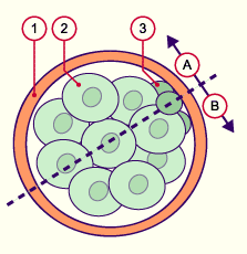
1
2
3
|
Pellucid zone
Blastomere
Polar bodies |
|
|
|
Fig. 17
Schematic sketch of an embryo in the morula stage. An imaginary equatorial plane goes through the collection of the polar bodies (dotted line). One pole of the embryo is on one side (A) and the other pole is on the other side (B).
|
|
|