|

|
|
|
Abnormalities in the closure of the neural tube
|
|
|
|
The largest portion of spinal cord abnormalities can be traced back to an unsuccessful closure of the neural tube, caudal to the 4th somite pair during the fourth week. This defect is termed spinal dysraphia and it influences not only the development of the central nervous system but also that of the vertebral arches that lie above it. This is evidenced by a more or less strongly pronounced opening of the vertebral canal (spina bifida). Transplantation experiments have actually shown that the neural tube exercises an inductive effect on the development of the vertebral arches 5.
According to the number of non-fused neural arches and of the other structures thereby affected, one distinguishes among various types of spina bifida.The clinical consequences can be insignificant, serious or even fatal.
Common to all types of spina bifida is an absent closure of the vertebral arch.
Basically one distinguishes between:
- Spina bifida occulta (= hidden)
- Spina bifida aperta (= visibly present)
- Myelocele
- Myelomeningocele - with or without cysts
- Myeloschisis
|
|
|
|
More info
|
|
Factors that could hinder neural tube closure:
The reasons for the malformation of the neural tube have yet to be found. One knows, though, that they must be multi-factorial. Genetic, as well as environmental and nutritional factors appear to play an important role. Studies have shown that the consumption of vitamins and folic acid can prevent the occurrence of the abnormality. Certain medications, though, for example valproic acid (an anti-epileptic medication) can increase the susceptibility to develop spina bifida.
|
|
|
|
|
More info
|
|
In order to find out more about the prevention of spina bifida using folic acid, consult this website (French).
|
|
|
|
Spina bifida occulta occurs very frequently and is usually found accidentally in x-rays or in an examination of the back. They seldom have clinical relevance because only a missing closure of the osseous structures exists in the formation of the vertebral arch, without the spinal cord with its membranes (meninges) being involved. The skin covering is intact. Sometimes a tuft of hair tells where the osseous structures are missing.
|
|
|
Fig. 29 - Spina bifida occulta
(longitudinal section) |
|
Fig. 29a - Spina bifida occulta
(cross section at the level of A) |
|
Legend |
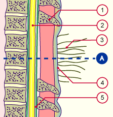
1
2
3
4
5 |
Spinous process
Spinal cord
Tuft of hair
Skin
Dura mater |
|
|
|
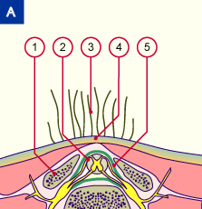
1
2
3
4
5
|
Vertebral arch
Spinal cord
Tuft of hair
Skin
Dura mater |
|
|
|
Fig. 29; Fig. 29a
Schematic drawing of a spina bifida occulta.
The meninges and the spinal cord are in their right places.
Optically, only a tuft of hair gives its presence away.
|
|
Spina bifida in combination with a dermoid cyst
|
|
|
It can happen that in the median, sacral region a fistula stays connected with a dermoid cyst, which indicates the location of the closure of the posterior neuropore in the 4th week. The cyst represents the last place of separation between the superficial ectoderm and the neuroectoderm. Spinal cord and meninges find themselves in their normal locations below the skin.
|
|
|
|
|