7.3 Pathologies


Spina bifida aperta


In cases of spina bifida aperta, besides fissures of the osseous structures, one also finds abnormalities of the meninges and/or of the spinal cord itself.
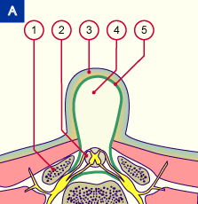
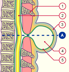
The membranous covering can be present or absent. The mildest form is the meningocele where merely the meninges protrude under the skin through the cleft in the malformed vertebral arch.


Fig. 30 - Meningocele
(longitudinal section)
Fig. 30a - Meningocele
(cross section at the level of A)
 Legend

1
2
3

4

5
Spinous process
Spinal cord
Mostly intact skin over the
meningocele
Meningocele with cerebrospinal
fluid
Dura mater


4
2
3

4

5
Vertebral arch
Spinal cord
Mostly intact skin over the
meningocele
Meningocele with cerebrospinal
fluid
Dura mater

Fig. 30; Fig. 30a
Schematic drawing of a meningocele. The spinal cord is in its normal location. Only a sack filled with cerebrospinal fluid, bounded by the dura mater, protrudes from below the skin.


In a myelomeningocele the meninges as well as the spinal cord (myelon) are found outside of the vertebral arch. They are visible as a protrusion under the skin; sometimes the skin can also be absent or only very thin.
In addition, the central canal is bloated and forms a cyst. In this case one speaks of a myelocystomeningocele.


Fig. 31 - Myelomeningocele
(longitudinal section)
Fig. 31a - Myelomeningocele
(cross section at the level of A)
 Legend

1
2
3

4
5
Spinous process
Central canal
Skin partially covering the neural
tube defect
Spinal cord
Dura mater


4
2
3

4
5
Vertebral arch
Central canal
Skin partially covering the neural
tube defect
Spinal cord
Dura mater

Fig. 31; Fig. 31a
Schematic drawing of a myelo-
meningocele.
The spinal cord comes to the surface through the osseous defect and is normally covered only by a thin skin.


More info

Macroscopic pictures


The myeloschisis (rachischisis) is the severest form of the spina bifida aperta. The nerve tissue is here fully bare and a dermal or meningeal covering is absent. With this abnormality, the closure of the neural folds fails to occur.


Abb. 32 - Myeloschisis
(longitudinal section)
Abb. 32a - Myeloschisis
(cross section at the level of A)
 Legende

1
2
3
4
Spinous process
Spinal cord
Dura mater
Skin up to the neural tube defect


4
2
3
4
Vertebral arch
Spinal cord
Dura mater
Skin up to the neural tube defect

Abb. 32; Abb. 32a
Schematic diagram of a myeloschisis.
The nerve tissue lies open at the surface without a dermal covering.
This leads to the severest disorder of the innervation.


Anencephaly


Anencephaly represents a special kind of spina bifida. It arises when the neural folds do not fuse in the cranial region of the neural tube. When the fusion fails for the entire neural tube, one speaks of craniorachischisis totalis. Both anomalies are always fatal.

More info

Macroscopic pictures


Beginning of the chapter | Previous page | Next chapter