|

|
|
|
|
The embryonic time comprises 56 days, i.e., 8 weeks from the moment of fertilization. This time span is divided into 23 Carnegie stages and the stage classification is based solely on morphologic features. Carnegie stages are thus neither directly dependent on the chronological age nor on the size of the embryo. This can be illustrated by two examples: The closure of the rostral neuropore occurs by definition in stage 11 and that of the caudal neuropore in stage 12. Further, between the 25th and 32nd days of the pregnancy, the stages are determined according to the number of the somites  9-13 9-13 that have been engendered. The individual stages thus differ in how long they last . that have been engendered. The individual stages thus differ in how long they last .
During the embryonic period most of the organ systems are established and this with an enormous rapidity. Cell divisions, movement and differentiation are the basic processes taking place during this phase. It is thus hardly surprising that this pregnancy phase is very vulnerable and that deformities are produced most often during this time. The type of deformity depends on the embryonic developmental stage.
|
|
|
| Fig. 1 - Incidence of deformities during pregnancy |
|
Legend |
A
B
|
Embryonic period
Fetal period |
|
|
|
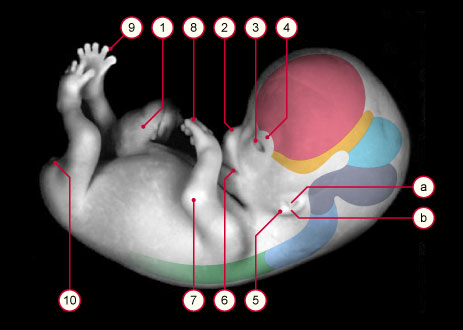
Fig. 1
Segment A represents the embryonic period in which the embryo is especially sensitive with respect to deformities.
Within the first eight weeks, the incidence of deformities (blue curve), that lead to miscarriages, decreases from more than 10% to 1% during the fetal period (B). The frequency of neural tube defects decreases from 2.5% to 0.1% (green curve) by the end of the embryonic period. (2)
|
|
According to estimates, over 90% of the 4500 designated structures of the adult body are already established - and can be distinguished - during the embryonic period (1). During the fetal period the organs that formed during the embryonic period grow and differentiate (organogenesis).
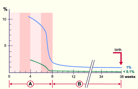
Figure 2 shows the various temporal phases during a pregnancy. A rough classification is made by assigning trimesters (trimenon). The LMP (Last Menstruation Period) is not the real beginning of the pregnancy but serves as a point of reference for determining the date of ovulation and thus the moment of fertilization. Normally this occurs 14 days after the beginning of menstruation, but can vary a lot temporally. From the time of the last period, one estimates 40 weeks after the last menstruation in order to determine the approximate date of birth (the second and third grid marks represent the lunar month [of 28 days] or 4 weeks). On average, though, the duration of an actual pregnancy amounts to 266 days or 38 weeks (fourth grid). The embryonic period (A) lasts 8 weeks and the fetal period (B) from the 9th week to the birth.
|
|
|
| Fig. 2 - Time calculations during a pregnancy |
|
Legend |
A
B
|
Embryonic period
Fetal period |
|
|
|
Fig. 2
The schematic diagram shows the various time periods during the entire pregnancy.
LMP = Last Menstruation Period.
The embryonic period (A) lasts 8 weeks and the fetal period (B) from the 9th week to the birth, i.e., 30 weeks.
|
|
In obstetrics the pregnancy weeks (PW) are normally reckoned from the date of the Last Menstrual Period (LMP). This is a point in time that many women can easily remember. Computed this way, the pregnancy lasts 40 weeks and the embryonic period - accordingly - 10 weeks. Caution is advisable, though, when wishing to calculate the moment of ovulation - and thus fertilization, closely connected with it - because the moment of ovulation can vary and depends on many factors (conditioned by the environment and psychological aspects). In embryology the temporal indices (i.e., the PW), therefore, always refer to the moment of fertilization even though in practical midwifery the time following the LMP is still used for computations.
After the 8th week, the fetus takes on typical human features, even though at the end of the first trimenon, the head is still relatively large in appearance. The eyes shift to the front and the ears and nasal saddle are formed.
The eyelids are also clearly recognizable now. On the body, fine lanugo hairs are formed, which at the time of birth are replaced by terminal hairs. The physiologic umbilical hernia that arises in the embryonic period 15-20 has mostly disappeared. In the second trimenon the mother feels the first movements of the child. In the last trimenon the subcutaneous fatty tissue is formed and stretches the still wrinkled skin of the fetus. The skin becomes covered more and more with vernix caseosa. This is a whitish, greasy substance und consists of flaked off epithelial cells and sebaceous gland secretions. In neonatology this vernix caseosa is an important criterion for judging the maturity of the child. If the birth occurs post-term, it disappears again.
|
|
|
|
|