|

|
|
|
Heart abnormality with left-right shunt (acyanotic)
|
|
|
|
With a left-right shunt an increased pulmonary perfusion to the detriment of the systemic circulation system is the result. Shunts from the oxygen-rich side to the oxygen-poor side are not usually accompanied by a cyanosis.
|
|
|
|
Persisting ductus arteriosus (PDA)
|
|
|
|
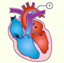
The ductus arteriosus connects the pulmonary artery with the aorta. Prenatal it is a vital structure to have. After birth, in the first days of life, though, the ductus arteriosus is closed by the active contractions of its smooth muscles, followed by an intima proliferation. The sealing of the ductus is triggered by the postnatal pO2 increase due to the breathing of the newborn.
|
|
|
| A persisting ductus arteriosus (PDA) is spoken of when the normal, postnatal closure fails to take place (ca. 9% of all cardiac abnormalities). In hemodynamic terms, the quantity of blood flow due to a PDA depends on the diameter and length of the ductus arteriosus. In addition, the size of this left-right shunt varies with the pulmonary resistance. Since in the first 3-8 weeks after birth this resistance decreases continuously, a cardiac insufficiency may occur. |
|
Fig. 36 - Persisting ductus arteriosus |
|
Legend |
|

|
|
Fig. 36
Due to the increase of the peripheral pressure in the systemic circulation system and the continuous lowering of the pulmonary resistance in the first weeks of life, a reversal of the flow through the ductus arteriosus and a flooding of the pulmonary vessels takes place.
|
|
More info
|
|
The symptoms of a persisting ductus arteriosus differ according to age:
- In infants with a large DA increasing cardiac insufficiency with tachypnea, dyspnea, and growth disorders are observed. Drinking weakness, accompanied by increased sweating occur when nursing because this represents a big effort for the newborn.
- In premature infants, an open DA leads to a flooding of the pulmonary circulation system.
- In older children an open DA is often discovered accidentally. It seldom leads to a symptom such as infection proneness.
Overview, according to the clinical picture, of the diagnostic possibilities as well as the therapy.
|
|
|
|
|
|
Atrial septum defect (ASD)
|
|
|
|
Prenatally an ASD is of no hemodynamic importance because the foramen ovale is already open normally and directs the blood from the inferior vena cava to the left side of the heart. Only after birth does it have hemodynamic consequences in that a left-right shunt arises due to the different pressures in the two atria. Its incidence amounts to 11%.
|
|
|
More info
|
|
One distinguishes according to localization among the following defects.
|
|
|
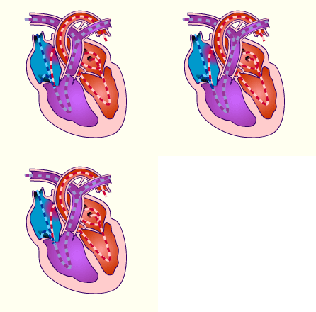
| The ASD leads to a left-right shunt and to an overload for the right ventricle with recirculation through the lungs.
|
|
Fig. 37 - Blood flow with an ASD |
|
Legend |
|

|
|
Fig. 37
The right circulation system is overloaded because blood from the left side always recirculates through the lungs via the ASD.
|
|
Ventricle septum defect (VSD)
|
|
|
|
Ventricular septum defects are encountered relatively frequently (28% of all congenital cardiac abnormalities). They can occur by themselves but also combined with other defects.
|
|
|
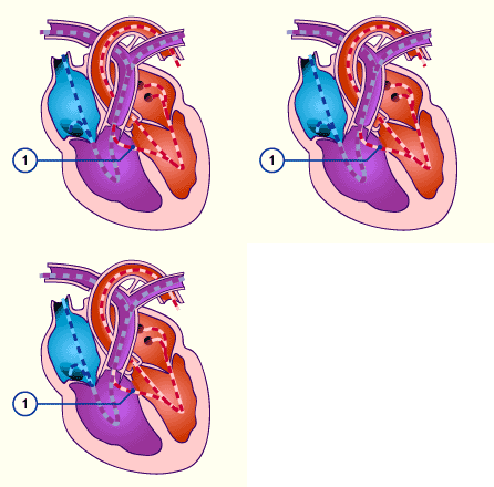
Prenatally, even a large defect is not disadvantageous because roughly the same pressure is present in both ventricles. Postnatally, the severity of the VSD depends on its size and the resistance relationships in the pulmonary and systemic circulation systems. Small defects lead to no detriment and especially those in the muscular septum sometimes close spontaneously.
Overview, according to the clinical picture, of the diagnostic possibilities as well as the therapy. |
|
Fig. 38 - VSD in the membranous part |
|
Legend |
|

1
|
Defect in the membranous part of the interventricular septum |
|
|
|
Fig. 38
In the membranous part a defect is present via which, in the absence of additional abnormalities, a left-right shunt from the left ventricle to the right exists.
|
|
Atrioventricular septum defect
|
|
|
|
|

