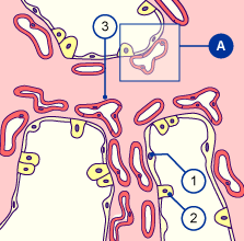
1
2
3
|
Type I pneumocyte
Type II pneumocyte
Capillaries |
|
|
|
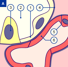
1
2
3
4
5
6 |
Type I pneumocyte
Saccular space
Type II pneumocyte
Basal membrane of the air passage
Basal membrane of the capillaries
Endothelium of the capillaries |
|
|
|
Fig. 7
The capillaries multiply around the acini. They push close to the surface and form a common basal membrane with that of the epithelium.
Fig. 8
The blood-air barrier in the lungs is reduced to three, thin layers: type I pneumocyte, fusioned basal membrane, and endothelium of the capillary.
|