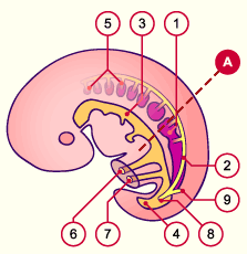
The mesonephric duct forms on the dorsal side of the nephrogenic cord at the level of the 9th somite. Initially it consists of a solid mesenchymal cord of cells  11 11 . It releases itself from the nephrogenic cord and is finally localized under the ectoderm, which probably plays an inductive role in its formation (3). . It releases itself from the nephrogenic cord and is finally localized under the ectoderm, which probably plays an inductive role in its formation (3).
Released from the nephrogenic cord, it develops in the caudal direction and canalizes itself at the same time  12 12 , in order to finally end in the cloaca. As soon as it is canalized one calls it the mesonephric duct (Wolffian duct). At the site where the mesonephric duct (Wolffian duct) discharges into the cloaca, the rear wall of the bladder forms. , in order to finally end in the cloaca. As soon as it is canalized one calls it the mesonephric duct (Wolffian duct). At the site where the mesonephric duct (Wolffian duct) discharges into the cloaca, the rear wall of the bladder forms. |
|
Fig. 6 - Development of the
mesonephros |
|
Legend |
|

1
2
1+2
3
4
5
6
7
8
9 |
Nephrogenic cord
Mesonephric duct
Mesonephros
Intestine
Cloaca
Atrophied nephrotome
Yolk sac (umbilical vesicle)
Allantois
Outflow of the mesonephric duct
into the cloaca
Ureter bud (anlage) |
|
|
|
Fig. 6
Sagittal section of a 5-week-old embryo. The pronephros atrophies while the mesonephric duct grows caudally and fuses with the cloaca wall. In this stage, it goes through a mesenchymal- epithelial transformation and forms a central lumen. Only the caudal part remains mesenchymal. Observe the ureter bud at its caudal end.
|

