20.5 Pathology


Congenital ureteral abnormalities


Course anomalies of the ureter


Retrocaval / retroiliac ureter:
In this abnormality the right ureter traces out an "S" at the L4 level behind the vena cava (retrocaval ureter). Responsible is a developmental disorder of the inferior vena cava. In a similar fashion the ureter can also run behind the common iliac artery at the L5 level (retroiliac ureter).
Fig. 37 - Retrocaval / retroiliac ureter


Anomalies of the ureteral diameter


Primary megaloureter due to an obstruction:
The cause of this abnormality is a constriction in the terminal part of the ureter, leading to a dilatation. The constriction can be anatomic (anomaly of the muscle layer) or of a purely functional kind (normal histology). Above the constriction the ureter is dilated, bulging and the wall is thickened. The primary megaloureter must be distinguished from the secondary that is due to a valve or a vesico-ureteral reflux.
Fig. 38 - Primary megaloureter due to obstruction

Macroscopy

Macroscopic picture


Quiz

Quiz 18


Abnormal number of ureters


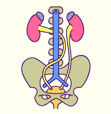
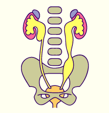
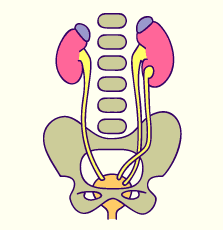
Disorders in the number of ureters belong to the most frequent anomalies of the urinary tract, whereby they are often asymptomatic. They arise from a premature branching of the ureter anlage, leading to a partial second or from an additional ureter anlage whereby, in this case, two complete ureters are generated.


Partial doubling:
Here an incomplete division of two ureters that fuse again at different levels (in the pelvis, lumbar, iliac or intramural regions). In every case a common end section exists with a normal ureteral orifice. A uretero-ureteral reflux often occurs at the level of the Y-branch.
Fig. 39 - Partial doubling


Complete doubling:
Here a complete doubling of the ureters with a second renal pelvis is involved. The ureters empty into the bladder in accordance with Weigert and Meyer's law (the upper ureter has its orifice lower down on the bladder than the lower one). The calyx system of the upper renal pelvis is not as well differentiated as that of the lower one. This abnormality often remains asymptomatic, except for complications that can arise due to the calyx system abnormalities.
Fig. 40 - Complete doubling

Macroscopy

Macroscopic picture


Previous page | Next page