|

|
|
|
21.5 The external genitalia
|
|
|
Differentiated stage of the male genitalia
|
|
|
|
Under the effects of androgen the differentiation and development of the male sexual organs become visible after the 3rd month.
The genital tubercle becomes longer and out of it forms the penis. The urethral folds also lengthen ventrally. Between these extends the urogenital sinus and forms the urethral groove, which is lined with endoderm. The floor of this sulcus thickens through epithelial proliferation and forms the urethral plate that temporarily fills it out.
Later a groove forms again and the two urethral folds fuse on the underside. This section will become the spongy part of the urethra, which for now terminates in a dead end in the anterior part of the penis.
In the rear part, the genital swellings transform themselves into the scrotum.
|
|
|
From the fused urethral folds an erectile mesenchymatous tissue, the penile spongy body, arises in the penis. At the distal penis section a ring-shaped furrow delimits the glans. Above the spongy body arise the two cavernous bodies (corpora cavernosa) and thus complete the penile erectile system.
The two genital swellings also fuse in the middle and form the scrotum. The line along which they fuse on the penis and scrotum is called the raphe mediana. |
|
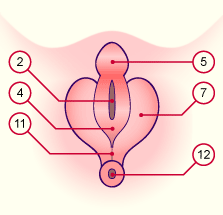
Fig. 57 - Differentiated external male
genitalia, ca. 10th week |
|
Legend |
|
|
|
Fig. 57
Lengthening of the genital tubercule and formation of the penile anlage. Proliferation on the floor of the urethral groove that is bounded by the urogenital folds.
|
Fig. 58 - Differentiated external male
genitalia, ca. 12th week |
|
Fig. 59 - Differentiated external male
genitalia, ca. 14th week |
|
Legend |

2
4
5
7
8
|
Urethral groove
Fused urethral folds
Penis
Genital swelling
Scrotum |
|
|
|
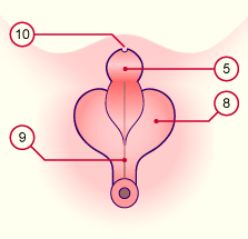
9
10
11
12
|
Raphe mediana (ano-genital)
Urinary meatus
Perineum
Anus |
|
|
|
Fig. 58
Increasing fusion of the urehtral folds at the ventral edge of the penis, back to front. This fusion forms the definitive spongy part of the urethra, which ends blindly shortly before the end of the penis.
Fig. 59
The two genital folds also fuse in the middle and form the scrotum. The line along the scrotum and penis where they fuse is called the raphe mediana.
|
|
During the 4th month two ectodermal invaginations arise on the tip of the penis. First, a solid epithelial cord forms from the penis tip and binds itself with the dead-ended spongy part of the urethra at the level of the ring-shaped furrow. As soon as this epithelial cord has been canalized, one speaks in this section of the glandar urethra with the urinary meatus.
The two circular epithelial ingrowths, glandar lamella, form the prepuce that at the time of birth is still stuck to the glans but, during childhood, comes away from it.
|
|
|
Fig. 60 - Penis, prepuce, spongy part
of the urethra, ca. 12th week |
|
Fig. 61 - Penis, prepuce, spongy part
of the urethra, ca. 14th week |
|
Legend |
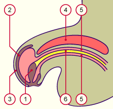
1
2
3
4
5
6
|
Epithelial hollow of the glans penis
Prepuce epithelial depression
Prepuce
Cavernous body
Spongy body
Spongy part of the urethra |
|
|
|
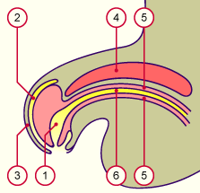
1
2 |
Glandar part of the urethra with
the navicular fossa
Resorbed prepuce epithelial
ingrowth |
|
|
|
Fig. 60
In the spongy part of the urethra forms erectile tissue, the penile spongy body, which extends into the glans penis. Above it the two cavernous bodies complete the penile erectile system.
Fig. 61
From the 14th week the two epithelial hollows on the glans penis open and create a connection within the glans between the spongy and glandar part of the urethra
|
|
|