|

|
|
|
21.1 Genetic factors and hormones that determine gender
|
|
|
Sertoli's supporting cells: function, mode of operation and hormonal secretion
|
|
|
|
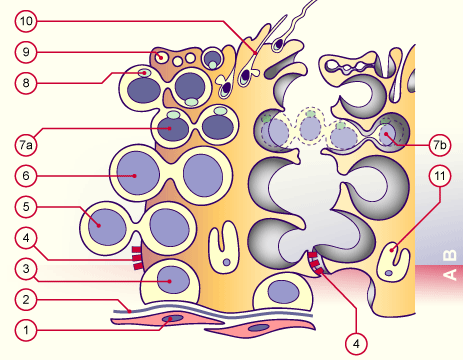
The supporting cells (Sertoli) are located within the seminiferous tubules. Their task is the creation of a hemato-testicular barrier and the nourishment of the spermatozoa. They can only proliferate in the first year of life (their equivalent in the woman are the follicular cells).
Sertoli cells are Christmas-tree-shaped and sit on a basal membrane. Laterally they stand in direct contact with one another and with the germ cells. The oval cell nucleus is indented and oriented perpendicular to the basal membrane and the nucleolus is clearly visible. These characteristics enable us to distinguish them from the germ cells.
Each supporting cell (Sertoli) is bound together with the neighboring cell through "tight junctions". This divides the germ epithelium into basal and adluminal compartments.
- The basal compartment contains spermatogonia up to the preleptotene stage (doubled DNA before the 1st meiosis).
- In the adluminal compartment are the spermatocytes, spermatids and spermatozoa. The "tight-junction" as a blood-testicle-barrier keeps spermatozoa from getting into the blood circulation or the lymphatic systems. This is important because the immune system would produce antibodies against the antigens on the membrane of the monoploid spermatozoa, leading to an autoimmune-orchitis and thus to sterility.
|
|
|
More info
|
|
Histology of the seminiferous tubule
|
|
|
| Fig. 2 - Supporting cells (Sertoli) (germinal epithelium |
|
Legend |

1
2
3
4
5
6
7a
7b
8
9
10
11
A
B |
Peritubular cells
Basal membrane
Spermatogonia
Tight junction
Spermatocyte I
Spermatocyte II
Spermatid
Spermatid
Acrosom
Residual bodies
Sperm cells
Nucleus of the supporting cells (Sertoli)
Basal Zone
Adluminal Zone |
|
|
|
Fig. 2
Schematic architecture of the germinal epithelium:
The supporting cells (Sertoli) sit on the basal membrane. Towards the lumen they are bound together through tight junctions. This seal engenders the blood-testicle-
barrier. The cytoplasm of these supporting cells gets formed into complicated processes, because they envelop all of the cells involved in spermatogenesis.
|
|
The function of the supporting cells (Sertoli) is controlled by the FSH pituitary hormone (follicle-stimulating hormone). Sertoli cells synthetize ca. 60 various proteins that are connected with reproduction. The most important are inhibin, androgen-binding-protein (ABP) and the antimüllerian hormone (AMH).
Antimüllerian hormone (AMH) is a glycoprotein of 560 amino acids and, together with inhibin, belongs in the TGF-b family. It is responsible for the atrophy of the paramesonephric duct (Müller) in men. The associated gene is localized on chromosome 19. The hormone can be detected very early in the developmental phase of the testicular differentiation and attains its maximum during the atrophy of the paramesonephric duct (Müller). In the "Persistent Müllerian Duct Syndrome" (PMDS) the paramesonephric duct (Müller) persists in a man that otherwise shows normal internal and external genitals. The reason is either a structural anomaly or a deficiency of AMH or its receptors. With the inception of puberty the amounts of AMH decrease greatly because testosterone, which is produced increasingly, inhibits the gene expression of AMH.
Inhibin is also a glycoprotein that inhibits the secretion of FSH. Inhibin is released in varying amounts in concert with testosterone.
|
|
|
|
ABP is a protein with a great affinity to testosterone and dihydrotestosterone. When it is released into the lumen of the seminiferous tubules, a concentration of androgens can be attained there that far exceeds its normal solubility. ABP is released under the influence of FSH and testosterone.
|
|
|
|
|

