10.2 Development of the placental villi


Anatomy of the placenta

Quiz

Quiz 04


In order to understand the chronological development of the chorionic villi it is important to have a comprehensive overview of placental anatomy. In this diagram, the placenta is roughly four months old and various fundamental structures can be recognized, namely the umbilical cord, the amnion, the chorionic plate, the already advanced branching of the villi, the basal plate and the cotyledon.


Fig. 13 - Placenta at around the fourth month  Legend

1
2
3
4
5
6
7
Umbilical cord
Amnion
Chorionic plate
Intervillous space (maternal blood)
Basal plate
Cotyledon
Villus

Fig. 13
Schematic diagram of the placenta at around the fourth month in a sagittal section.


Note: Zone A corresponds to the interactive diagram of this module


At birth, the placenta consists of two parts:

  • maternal portion
  • fetal portion


Fig. 14 - maternal side Fig. 15 - fetal side  Legend

1
2
Cotyledon
Cut edge of the amnion


3
4
Umbilical cord
Decidua with the compact layer after the release of the placenta

Fig. 14
Placenta: View from the maternal side
Fig. 15
Placenta: view from the fetal side


Fig. 16 - Placenta: the maternal side Fig. 17 - Placenta: the fetal side  Legend

1
2
Cotyledon
Cut edge of the amnion


3
Umbilical cord

Fig. 16
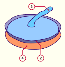
From the maternal side the placenta has a wrinkled surface, characterized by the cotyledons.
Fig. 17
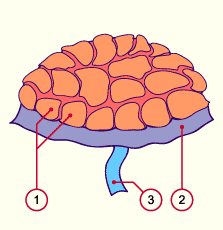
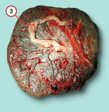
From the fetal side, the placenta is smooth and shiny. Notice the normal, central insertion of the umbilical cord in this case.

© Institut de pathologie, CHUV, Lausanne


List of the chapters | Next page