|

|
|
|
10.2 Development of the placental villi
|
|
|
|
While the embryo is nourished in the first weeks through simple diffusion, later, due to its rapid growth, it needs a more powerful gas and nutrient exchange system. This is made possible by the development of the utero-placental circulation system in which the circulation systems of the mother and of the embryo get closer together, thus allowing an exchange of gases and metabolites via diffusion.
It must be always kept in mind, though, that maternal and fetal blood never come into direct contact with each other.
This system decays after the ninth day in the lacunar stage  5b 5b . .
Through the lytic activity of the syncytiotrophoblast (Fig. 18 and 19) the maternal capillaries are eroded and anastomose with the trophoblast lacunae, forming the sinusoids. At the end of the pregnancy the lacunae communicate with each other and form a single, connected system that is delimited by the syncytiotrophoblast and is termed the intervillous space.
|
|
|
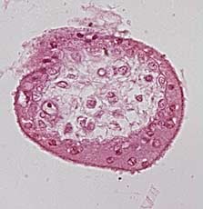
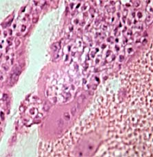
| Fig. 18: 9th .10th day - Lacunar stage |
|
Fig. 19: 9th -10th day – Primary villus |
|
Legend |
1
2
3
4
|
Cytotrophoblast
Syncytiotrophoblast
Spaces between
syncytiotrophoblast (Lacunae)
Maternal vessel |
|
|
|
5
A |
Maternal vessel, eroded by the ST,
which form the maternal sinusoids
through communication with the
lacunae
See enlarged version in figure 20 |
|
|
|
Lacunar stage (Fig. 18) and primary villus (Fig. 19)
Spaces form in the trophoblast (Fig. 18). Subsequently, due to the erosion of the maternal capillaries, blood gets into the vacuoles, engendering the maternal sinusoids. (Fig. 19)
|
|
Between the 11th and 13th day cytotrophoblast cells penetrate into the cords of the syncytiotrophoblast creating the primary trophoblast villi 5b.
|
|
|
| Fig. 20: 11th -13th day |
|
11th -13th day |
|
Legend |
1
2
|
Cytotrophoblast
Syncytiotrophoblast |
|
|
|
|
|
Primary villus with the cytotrophoblast, which penetrates into the processes of the syncytiotrophoblast, forming the primary trophoblast villi.
|
|
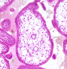
After the 16th day the extra-embryonic mesoblast also grows into this primary trophoblast villus, which is now called a secondary villus 5c and expands into the lacunae that are filled with maternal blood. As was already mentioned, the ST forms the outermost layer of every villus.
|
|
|
|
At the end of the 3rd week the villus mesoblast differentiates into connective tissue and blood vessels. They connect up with the embryonic blood vessels. Villi that contain differentiated blood vessels are called tertiary villi 6.
|
|
|
|
From this time on gases, nutrients, and waste products that diffuse through the maternal and fetal blood must pass through a total of four layers:
- Capillary endothelium of the villus
- Loose connective tissue that surrounds the endothelium
- Cytotrophoblast
- Syncytiotrophoblast
These four elements together form the placental barrier.
Note! The endothelium that surrounds the maternal blood vessels never penetrates into the trophoblast lacunae, but comes just to their boundaries.
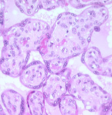
Numerous "daughter" villi arise out of the tertiary villi. These remain either free and project into the intervillous space (free villi), or they anchor themselves to the basal plate (anchoring villi). (Interactive diagram).
|
|
|
|
After the 4th month the cytotrophoblast in the tertiary villi disappear slowly, the villi divide further and become very thin, whereby the distance between the intervillous space with maternal blood and the fetal vessels gets smaller. The villi that arise in this way are called free villi.
|
|
|
|
|