10.2 Development of the placental villi


The cytotrophoblast layer


The cytotrophoblast of the anchoring villus expands until a further layer outside the syncytiotrophoblast arises, forming the cytotrophoblast layer (interactive diagram). It slips in between the syncytiotrophoblast and the uterine endometrium. Fig. 24 - Development of the cytotrophoblast layer  Legend

1
2
3
4
5
Anchoring villus
Syncytiotrophoblast
Cytotrophoblast
Cytotrophoblast layer
Uterine endometrium

To be recognized are the anchoring villus with the cytotrophoblast shown in dark green that infiltrates more and more the basal plate and gets in between the syncytiotrophoblast (light green) and the compact layer of the decidua in order to form the cytotrophoblast layer. Note that the syncytiotrophoblast completely covers the interior of the intervillous spaces.


Over the course of the 4th month the cytotrophoblast cells slowly disappear out of the villus wall and the chorionic plate. They persist, however, in the cytotrophoblast layer. The cytotrophoblast cells penetrate into the decidua and the myometrium and also colonize the wall of the spiral arteries close to their openings.


Fig. 25 - Growth of the CT into the walls of the maternal vessels Fig. 26 - Growth of the CT into the walls of the maternal vessels  Legend

1
2
3
4
Syncytiotrophoblast (ST)
Cytotrophoblast (CT)
Endothelial cells
Smooth muscle cells


5
6
Spiral arteries
Endovascular cytotrophoblast

Fig. 25, Fig. 26
Progressive growth of the cytotrophoblast cells into the decidua and into the wall of the spiral arteries.


This invasion of the maternal vessels by the cytotrophoblast leads to the destruction of the smooth muscle layer and to a partial replacement of the endothelial cells. It is responsible for the change in elasticity of the spiral arteries, whereby the blood circulation of this fetoplacental unit is adapted to the rapid growth of the fetus. This phenomenon of cell exchange is absent in preeclampsia or an intra-uterine growth retardation.An excessive proliferation of the cytotrophoblast can lead to tumor formation, especially to a chorion carcinoma.

More info

Supplementary information regarding the physiopathology of the mechanisms of preeclampsia

Physiopathology of preeclampsia (French)

HTA of the pregnancy (French)


More info

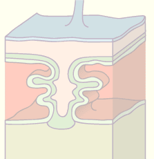
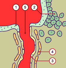
During a normal pregancy the maternal spiral arteries that nourish the placenta are continuosly pulled into the lacunar system.
These structural adaptations are accompanied by an edema, the dissolution of the endothelium and destruction of the tunica media and the membrana elastica interna, which are replaced by fibrous tissue. Through these alterations the arteries are removed from neuro-vascular control and the influence of the tone-producing vessel mediators (prostaglandin, nitrous oxide, endothelin). Thus, a larger blood flow is allowed in the placenta.
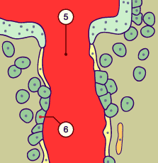
The migration of the trophoblast cells stands under a strict temporal-spatial control, an alteration of which can provoke a disorder in placental function. This ranges from a preeclampsia (characterized by insufficient penetration by the trophoblast) to a chorion carcinoma (characterized by an excessive trophoblast invasion).

Quiz

Quiz 16


Previous page | Next page