8.3 Folding of the germinal disk and the generation of the abdominal wall (4th week)


Introduction

Quiz

Quiz 04


After the end of the third week, the three germinal layers begin to differentiate and transform so that the initially flat embryonic disk develops into a cylindrical structure like a "C". The folding and genesis of the abdominal wall permits a delimitation of the embryo, that can now be clearly distinguished from the appending organs. Up to this stage 9 , the extraembryonic tissue went over into the intraembryonic tissue with no boundaries. The folding and the resulting formation of the abdominal wall lead to an enclosure of the mesoderm and the endoderm. They become surrounded by the ectoderm, which later forms the skin.

Two mechanisms lead simultaneously to the formation of the abdominal wall:

  • The cephalo-caudal flexion (in the longitudinal direction)
  • The lateral folding (in the transversal direction, rolling up)

Review

This sign points to a Carnegie stage.

Carnegie:
12. If you click on it, a pop-up window opens showing the embryo in stage 12. All further stages can also be seen in the windows.


The cephalo-caudal folding (in the longitudinal direction)

Quiz

Quiz 01


In order to understand how this turning takes place, the structures must first be described that are found in the cephalic end before the folding:
In the cephalic region, rostral to the prechordal plate and the pharyngeal membrane, the mesenchymal cells form the cardiac plate (pericardium) and the septum transversum (which later becomes a part of the diaphragm and separates the coelom into thoracic and abdominal cavities).
With the 180° degree turn that results from the folding, the following occurs:
the pharyngeal membrane extends towards the lower front (mouth area) and the cardiogenic plate (which initially lay most cranially) into the thorax area. Between the cardiac anlage and the umbilical vesicle a mesenchymal bridge forms, the septum transversum.
After this movement is completed, the brain (encephalon) lies the most cranially, followed by the mouth, heart, and diaphragm (septum transversum). During this folding the endoderm below the pharyngeal membrane becomes surrounded ventrally by the cardiac anlage. From this region, the throat (pharynx) arises and, subsequently, the thyroid gland, the lungs, and the esophagus. The pharyngeal membrane that for now separates the mouth (ectoderm) from the throat (endoderm) later atrophies 13.

What ?
How ? Where ?

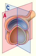
- Cephalo-caudal flexion takes place in the A plane

- Lateral folding takes place in the C plane


In order to understand the process of folding better (after stage 9), the forgoing stages are also depicted.


1
2
3
4a
4b

4c
5
6a
6b
7
8
9
10a
10b
11a
11b
12
13
14a
14b
14c
15
16
17
18
19
20
21
22
23
24
25
26
27
28
Amniotic cavity
Ectoblast
Umbilical vesicle
Endoblast
Endoblast that joins together
again under the notochord
Completely joined up endoblast
Body stalk
Allantois
Lengthened allantois
Extraembryonic mesoblast
Cloacal membrane
Notochordal process
Primitive streak
Shrinking primitive streak
Neural plate
Neural folds
Central axial canal
Primitive node
Ventrally merged notochord
Ventrally resorbed notochord
Free notochord
Prechordal plate
Canalis neurentericus
Pharyngeal membrane
Cardiac plate
Pericardial cavity
Fused neural tube
Septum transversum
Closure of the neuroporus cranialis
Recessus caudalis
Thyroid gland anlage
Lung anlage
Liver anlage
Closure of the neuroporus caudalis
Dorsal aorta
Longitudinal folding Carnegie
stages 6 - 12
Navigation


The gray elements from 29 to 37 are only visible in the transversal section. This is depicted on the following page.
29
30
31
32
33
34
35
36
37
Aorta
Umbilical veins
Intraembryonic mesoderm
Paraxial mesoderm
Intermediate mesoderm
Lateral plate mesoderm
Canalis centralis
Somite
Nephrogenic cord
without definition
with definition


stage 6
(17th day)

stage 7
(19th day)

stage 7
(21rst day)

stage 8
(23rd day)

stage 8
(24th day)

stage 9
(25th day)

stage 9
(27th day)

stage 10a
(28th day)

stage 10b
(28th day)

stage 10c
(28th day)

stage 11
(29th day)

stage 12
(30th day)


Animation of what is depicted in the above diagrams


Fig. 6 - Folding of the cephalic end
S9 (ca. 27th day)
Fig. 7 - Folding of the cephalic end
S12 (ca. 30th day)
 Legend

1
2
3
4
5
6
Future prosencephalon
Notochord
Neural tube
Pericardial cavity
Cardiac tube
Pharyngeal membrane


7
8
9
Extraembryonic mesoderm
Throat
Septum transversum

Fig. 6, 7
Schematic detail of the cephalic extremity, sagittal section


The folding of the caudal end occurs after the cephalic folding and has the result that the body stalk comes closer to the umbilical vesicle (yolk sac).
Due to the large axial growth the caudal end of the embryonic disk (with the cloacal membrane) comes to lie under the original embryonic disk and thus shoves the allantois and the body stalk in the ventral direction, up to the umbilical vesicle (yolk sac) and merges with its stalk.
To be noted is that now the end of the primitive streak, which initially lies dorsally after the flexion of the embryo, also comes to lie ventrally.


Fig. 8 - Folding of the caudal end
S9 (ca. 27th day)
Fig. 9 - Folding of the caudal end
S12 (ca. 30th day)
 Legend

1
2
3
4
5
6
7
8
Notochord
Neural fold
Amniotic cavity
Primitive streak
Primary endoderm
Cloacal membrane
Allantois
Body stalk


1
2
3
5
6
7
8
9
Notochord
Neural tube
Amniotic cavity
Endoderm
Cloacal membrane
Allantois
Body stalk
Hind gut

Fig. 8, 9
Schematic detail of the folding of the caudal end.


List of the chapters | Next page