8.3 Folding of the germinal disk and the generation of the abdominal wall (4th week)


Condition of the folding at the end of the 4th week


For a better understanding of the folding at the end of the fourth week 12 several regions have to be looked at:

  • The region above the umbilical cord (Fig. 9, section B)
  • The region in the area of the umbilical cord (Fig. 9, section C)
  • The region below the umbilical cord (Fig. 9, section D)


Fig. 9 - Schematic diagram of the finished folding,
the abdominal wall (ectoderm) has been formed (stage 12)
 Legend

Fig. 9
Transversal section B above the umbilical cord.

Transversal section C at the level of the umbilical cord.

Transversal section D below the umbilical cord.


In the region above the umbilical cord (Figs.10 - 11) the liver anlage (ventral) 11 is formed from the endoderm and somewhat later the dorsal pancreas anlage 12.

The liver diverticulum appears cranially to the umbilical vesicle (yolk sac). Liver cell bands penetrate into the mesoderm of the septum transversum. This is formed at the ventral location between the liver and cardiac anlage and contains the umbilical vein.


Fig. 10 - Region above the umbilical
cord (stage 11, ca. 29th day)
Fig. 11 - Region above the umbilical
cord (stage 12, ca. 30th day)
 Legend

1
2
3
4
5
Nephrogenic cord
Aortae
Dermatomyotoma
Ectoblast
Notochord


6
7
8
9

10
11
12
13

14
Pancreas anlage
Intraembryonic coelom
Amnion
Somatopleura mesoderm and ectoderm
Gut
Liver anlage
Septum transversum
Splanchnopleura mesoderm und endoderm
Amniotic cavity

Fig. 10
The ventral abdominal wall now closes itself. From the endoderm the liver anlage is formed ventrally. In stage 12 the pancreas anlage is visible.

Fig. 11
The ventral abdominal wall is closed. From the endoderm the pancreas anlage forms dorsally. The liver develops much further in this stage and partially fills the coelomic cavity.


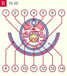
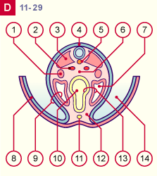
In the region below the umbilical cord (Fig. 13), the ventral abdominal wall closes up, whereby the primitive gut remains connected to it by the ventral mesenterium. After resorption of the ventral mesenterium, the gut is attached to the dorsal torso wall solely through the dorsal mesenterium.


Fig. 12 - Region below the umbilical
cord (stage 11, ca. 29th day)
Fig. 13 - Region below the umbilical
cord (stage 12, ca. 30th day)
 Legend

1
2

3
4
5
6
7
Nephrogenic cord
Aortae
(their fusion occurs in stage12)
Dermatomyotoma
Ectoblast
Notochorde
Mesenterium dorsal
Intraembryonic coelom


8
9

10
11
12
13

14
Amnion
Somatopleura mesoderm and
ectoderm
Gut
Allantois
Mesoderm
Splanchnopleura mesoderm and
endoderm
Amniotic cavity

Fig. 12
The embryo becomes totally enveloped by the amnion now. The intraembryonic coelom encloses the gut and temporarily forms a ventral mesenterium, that attaches the gut to the ventral abdominal wall.


Fig. 13
The ventral mesenterium has disappeared and, except for the umbilical cord, the embryo is completely surrounded by the amnion (later the peritoneal cavity will form at this level).


Beginning of the chapter | Previous page | Next chapter