|

|
|
|
14.3 Skeletal musculature
|
|
|
Differentiation of the somites
|
|
|
|
The tissue that immigrates via the primitive streak and initially forms the third germinal layer, the mesoderm, transforms itself several times.The epiblast-epithelial (ectoderm cells) are transformed into segmented mesoderm cells. In contrast to the epithelial cells, these are loosely organized. A new transformation follows, i.e., an epithelialization to become somites, which represent an epithelialized mesenchyma portion with a central somitocoel.
|
|
|
 |
|
|
|
|
 |
|
Legend
|
|
Think about what takes place between the various cell forms. For the answer click on  . .
|
|
|
|
|
|
|
|
These somites develop on both sides of the neural plate, caudal to the otic placode (post-otic), and are square-shaped, externally easily visible formations.
In their form as epithelialized somites, they do not remain very long, but rather very early exhibit a polarization in all directions.
|
|
|
| Fig. 11 - Transverse section through an embryo in stage 11, ca. 29 days |
|
Legend |

1
2
3
4
5
6
7
8
|
Somite
Somitocoel
Neural tube
Central canal of the neural tube
Coelom
Intermediary mesoderm
Splanchnopleura
Somatopleura |
|
|
|
Fig. 11
On the left is shown an embryo in stage 11, ca. 29 days.
|
More info
|
|
Detailed information about the structures in this diagram.
|
|
On the right a transverse section through the embryo at level A is displayed. The somites at this level exhibit a somitocoel. This is a lumen that is surrounded by the epithelialized cells of the somite.
|
More info
|
|
Detailed information about the structures in this diagram.
|
|
|
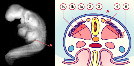
| Fig. 12 - Stage 12, ca. 30 days |
|
Legend |

1a
1b
1c
2
3
4
5
|
Dermatome
Myotome
Sclerotome
Central canal of the neural tube
Neural tube
Pars epaxialis
Pars hypaxialis |
|
|
|
Fig. 12
On the left is shown an embryo in stage 12, ca. 30 days.
On the right a transversal section through the embryo at level A is displayed. The somites have released themselves and form dermatomes, myotomes and sclerotomes. The aorta is no longer paired.
|
|
In the group of myoblasts that have a hypaxial origin and that have partially migrated into laterally lying regions of the embryo, the segmental ordering is no longer so clearly visible. It is interesting in this connection that over the course of the development the placement or definitive extension of a muscle can change. This happens, for example, in the muscles that affect the shoulders. The latissimus dorsi muscle and the major pectoral muscle obtain a secondary connection to the trunk part of the skeleton, but retain their original innervation from the brachial plexus.
|
|
|
|
More info
|
|
The myogenesis of the muscles occurs in several waves:
- The myotome development of the somatic epaxial muscles (5) takes place in several steps and is characterized by waves of myoblasts that emigrate, one after the other, to their future locations.One assumes that somite formation is multifactorial and regulated by various genes. Today, with the help of mutation analyses, one knows various genes (Fgfr1, Tbx6, Wnt 3a) that play important roles in somite formation. One assumes that such genes are mainly activated during the inflow of cells via the primitive streak and nodes. Nevertheless, one still does not know the precise mechanism that leads to metamerism. One can only guess that regional gene activities are responsible for somite formation. In the following interactive pars epaxialis diagram, (180 kB) the migration and differing gene activities of the epaxial musculature, depending on the developmental stage, can now be observed somewhat more precisely.
- The muscles that stem from the pars hypaxialis (interactive pars hypaxialis diagram, 165 kB) of the myotome (6), also develop dorsomedially to ventrolaterally in two waves. In contrast to the epaxial musculature few developed premyoblasts migrate into the periphery and only there develop into the masses of premuscles and into the definitive musculature.
|
|
|
|
|
|
|

