|

|
|
|
16.7 Development of the arteries
|
|
|
Vessels of the dorsal aorta
|
|
|
|
The right and left dorsal aortas (later descending aorta) develop in parallel with the heart and gain access to it via the aortic arches. In a lengthy process the paired dorsal aortic anlagen unite and finally form the unpaired descending aorta of the adult.  12-17 12-17
|
|
|
|
One can subdivide the various branches of the dorsal aorta into three groups:
- ventral (visceral), segmental branches
- lateral (visceral), segmental branches
- dorsolateral (parietal), intersegmental branches
These branchings are modified in various ways until they reach the definitive adult form.
|
|
|
|
Ventral (visceral), segmental branches of the dorsal aorta
|
|
|
|
The ventral segmental, visceral branches of the dorsal aorta, which initially appear paired and, in a large number, surround the intestines and the umbilical vesicle, from dorsal to ventral. With the further development of the intestines, in that the two layers of the mesenterium dorsale approach each other, the paired branches fuse in the middle and, from cranial to caudal, form the truncus coeliacus and the superior and inferior mesenteric arteries in that order.
The umbilical arteries are engendered from a vessel plexus in the lower part of the still paired dorsal aorta 10 at the level of the allantois. They are segmental arteries that soon gain access, though, to the intersegmental, lumbar arteries, whereby the cranial parts atrophy. The result is that these lumbar portions become the main part of the umbilical arteries. Parallel to the development of the subclavian artery of the arms the blood supply of the leg anlagen also develops from dorsolateral branches of the dorsal aorta. In that the umbilical arteries have incorporated the proximal portion of the dorsolateral branches, though, the leg arteries appear to spring from the umbilical artery.
In adults, the superior vesical artery and the median umbilical ligament (the obliterated intracorporal part of the umbilical artery) in the lower abdominal wall form the remnants of the arterial part of the umbilical circulation system.
|
|
|
Fig. 29 - Ventral branches (embryo)
Stage 19 (ca. the 46th day) |
|
Fig. 30 - Ventral branches
in adults |
|
Legend |
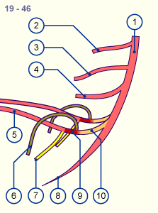
1
2
3
4
5
6
7
8
|
A. dorsalis
Truncus coeliacus
Superior mesenteric artery
Inferior mesenteric artery
Umbilical artery
(median umbilical ligament)
Ischiadic artery
Median sacral artery
Femoral artery |
|
|
|
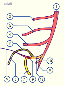
9
10
11
12
|
Umbilical artery (section of the
later internal iliac artery)
Umbilical artery (section of the
later common left iliac artery)
External iliac artery
Common right iliac artery |
|
|
|
Fig. 29
Overview of the various paired and unpaired branches of the dorsal aorta in the abdominal region. The individual sections of the embryonic vessels, which remain until adulthood, are shown in differing colors.
Fig. 30
In this picture the common left iliac artery (left umbilical artery) has been cut away to provide a better overview. The colors correspond to the sections of Fig. 29.
|
|
The median sacral artery originates at the branches of the two umbilical arteries. It also comes originally from the dorsolateral parietal branches of the aorta.
|
|
|
Summarizing, the displacement of the umbilical artery caudally results in the following situation:
- Common iliac artery: proximal part of the umbilical artery, beginning at the aortic branch.
- External iliac artery: proximal part of the leg artery, originally from dorsolateral branches but later from the umbilical artery.
- Internal iliac artery: continuation of the umbilical artery towards the navel after the branching off of the external iliac artery.
- Superior vesical artery: last vessel branching before the obliteration of the umbilical artery.
|
|
|
|
|
|
Lateral (visceral), segmental branches of the dorsal aorta
|
|
|
|
The second group consists of ca. 20 pairs of lateral visceral branches that are responsible for the blood supply of the mesonephros. From them paired branches come, namely the arteries for the suprarenal glands, the kidneys and the gonads.
|
|
|
|
Dorsolateral (parietal), intersegmental branches of the dorsal aorta
|
|
|
|
As the third group, the dorsolateral parietal branches form in the thoraco-lumbal part the intercostal and lumbar arteries and in the lowest part the median sacral artery which, after the various mentioned transformation processes, spring as unpaired arteries from the umbilical artery. In the cervical part, an anastomosis forms between the intersegmental cervical branches. From this long anastomosis arise on both sides the vertebral artery, which comes left and right from the subclavian artery. This is primarily a result of the transformation processes of the aortic arch arteries. The vertebral artery fuses to the basilar artery at the base of the brain and forms a blood supply source for it. The other supply sources of the head have already been treated in connection with the aortic arches and consist of the internal carotid artery for the anterior and lateral part of the brain and the external carotid artery for the face.
|
|
|
|
Summary of the dorsal aortic vessels
|
|
|
| Dorsolateral intersegmental branches |
| Dorsolateral intersegmental branches |
- Vertebral artery from anastomosis between the cervical intersegmental branches of the paired dorsal aorta
- Subclavian artery from the 6th cervical intersegmental artery dorsolateral branches (intersegmenal arteries), before the dorsal aortae fuse
- Intercostal arteries from the thoracic intersegmental arteries
- Iliac arteries partly from the lumbar intersegmenal arteries, partly through fusion with the umbilical artery
|
| Ventral segmental branches |
| Vessels of the umbilical vesicle |
- Truncus coeliacus
- Superior mesenteric artery from the omphalomesenteric artery
- Inferior mesenteric artery
|
| Vessels of the allantois |
|
| Lateral segmental branches |
| Lateral segmental branches |
- Medial suprarenal artery
- Renal artery
- Ovarian and testicular (spermatic) artery
|
|
|