|

|
|
|
16.7 Development of the arteries
|
|
|
|
The aortic arches are vessels that connect the cardiac saccus aorticus with the paired dorsal aorta by going around the pharynx. They develop one after the other in the pharyngeal arches. Between them the pharyngeal pouches are found that represent endodermal protrusions. When the aortic arches 3-6 have appeared  14 14 , the first two have already more or less disappeared. , the first two have already more or less disappeared.
It seems the neural crest cells, which migrate via the aortic arches to the heart, also have a large influence on the normal development of the aortic arches (compare separation of the outflow tract).
|
|
|
 |
|
|
|
|
|
| This successive appearance of the aortic arches reflects the principle of morphologic adaptation of the vascular system to the various stages of embryonic development. |
|
Fig. 28 - Connection of the
aorto-pulmonary septum
with the truncus septa |
|
Legend |
|
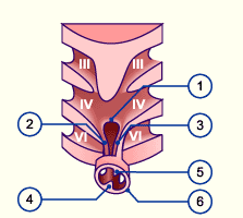
1
2
3
4
5
6 |
Aorto-pulmonary septum
Right aorto-pulmonary septum
pedicle
Left aorto-pulmonalry septum
pedicle
Upper truncus process
Lower truncus process
Truncus |
|
|
|
Fig. 28
The aorto-pulmonary septum grows upstream from material between pharyngeal arches IV and VI. It connects up with the two truncus processes. By clicking the relationships with the associated aortic arch arteries can be visualized. |
- The first 3 arches were adapted for supplying the cervical and cranial regions and, among other things, form the internal carotid artery 11, which is responsible for the supplying the face and frontal parts of the brain.
The external carotid artery 14 arises somewhat later as an independent vessel.
- With the asymmetric development of the heart and the division of the outflow tract, the 4th aortic arch also develops asymmetrically. The left 4th aortic arch remains as the arcus aortae of the adult while the right one forms the proximal part of the right subclavian artery.
- 6 aortic arches have always been spoken about. The last two, however, never appear in a prominent arch-shape like the first 4. The 5th aortic arch forms only a small capillary network and the 6th appears as a prominent capillary network with the early development of the trachea and lungs. It is also called the pulmonary arch. Only its left dorsal branching from the dorsal aorta forms a proper vessel, the ductus arteriosus. The ductus arteriosus via its connection with the left ventral branching of the saccus aortae and truncus pulmonalis, forms a shunt from the lung vessels to the dorsal aorta. In this way a too forceful perfusion of the still tender pulmonary capillaries by the blood from the right ventricle is avoided.
- The left subclavian artery arises from the left intersegmental 6th artery in the region of the 6th -7th cervical segment. Through differing growth processes (compare descent of the heart) it appears as if the left subclavian artery arises directly from the aorta. In the literature differing opinions about the origin of the left subclavian artery can be found. Some authors describe it as a derivative of the 6th, other of the 7th intersegmenatl arteries. (14)
|
|
|
More info
|
|
For the development of the ductus arteriosus the expression of Hoxb5 by the neural crest cells along the 6th aortic arch is probably determinant. The ductus arteriosus is a vessel of the muscular type. It is held open prenatally through the influence of prostaglandines that stem from the placenta, and through the relatively low partial pressure of the oxygen (pO2). (compare adaptation at birth)
|
|
|
|
More info
|
|
The detailed depiction of the aortic arch arteries is intended to show how the asymmetry of the vessels arise in the arcus aortae region.
|
|
|
|
|
|
Overview of the origin of the arteries that stem from the aortic arch arteries.
|
|
|
|
|