|
The atrioventricular conduction system, the av-node or Aschoff-Tawara's node, becomes discernible somewhat later
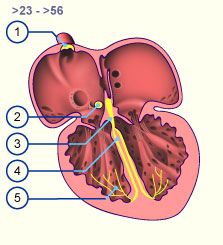
 15-16 15-16 . It lies at the dorsal circumference of the av-canal in the inner layer of the myocardium at the beginning of the dorsal av-septum. From it derives the His' bundle, which extends over the dorsal av-septum and forms the connection between the atrial and ventricular myocardium Here it divides into three subendocardial bundle branches. They extend into the cardiac apex region where a separation into fine Purkinje's fibers occurs. In situ, this conduction system forms itself from specialized myocardial cells. Once the sinu-atrial node . It lies at the dorsal circumference of the av-canal in the inner layer of the myocardium at the beginning of the dorsal av-septum. From it derives the His' bundle, which extends over the dorsal av-septum and forms the connection between the atrial and ventricular myocardium Here it divides into three subendocardial bundle branches. They extend into the cardiac apex region where a separation into fine Purkinje's fibers occurs. In situ, this conduction system forms itself from specialized myocardial cells. Once the sinu-atrial node  14-16 14-16 as well as the av-node and His' bundle have been differentiated, the cardiac frequency increases rapidly and reaches 140 beats/min. The region with the highest frequency (sinus region) takes over the pacemaker function. as well as the av-node and His' bundle have been differentiated, the cardiac frequency increases rapidly and reaches 140 beats/min. The region with the highest frequency (sinus region) takes over the pacemaker function.
|
|
Fig. 19 - Conduction system of
the embryonic heart |
|
Legend |
|

1
2
3
4
5
|
Sinu-atrial node in the sulcus
terminalis
Av-node (Aschhoff-Tawara node)
Truncus (His' bundle)
Right and left pedicle bundle
Purkinje fibers
|
|
|
|
Fig. 19
The pacemaker for normal heart rhythm is the sinus node. The excitation spreads out over the two atria and extends to the av-node, which also possesses the pacemaker capability. From here, the excitation spreads over the His' bundle and bundle branches and finally via the Purkinje fibers into the ventricular myocardium.
|

