|

|
|
|
16.1 Outer form and position of the heart
|
|
|
|
In the young embryo epiblast cells wander via the primitive streak between epi- and hypoblast and form the mesoderm layer out of which various structures arise. Cells, which are destined for cardiogenesis, take up a position that is cranial to the forming neural tube. Cardiogenesis takes place via a complex series of steps:
- Determination of mesoderm- and neural crest cells for heart formation
- Growth and differentiation processes to become cardiomyocytes
- Migration and transformation processes in order to form the heart
|
|
|
First signs of cardiac development
|
|
|
|
The cardiogenic plate is formed by a collection of mesoderm cells in the most anterior part of the embryo.
In the interactive diagram, please observe how the position of the pericardial cavity changes in relation to the cardiogenic plate due to the bending of the cranial end of the embryo.
|
|
|
|
The blood from the supplying vessels, the umbilical and omphalomesenteric veins, flows caudally over the inflow tract into the cardiac anlage and leaves it via the outflow tract and the aortic arches at the cranial end.
|
|
|
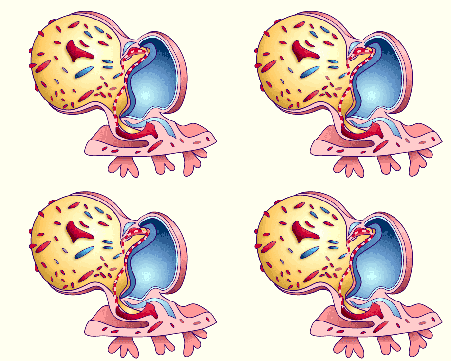
Fig. 1 - Embryo at stage 9 (roughly 25 - 27 day)
after the rotation of the cardiac anlage |
|
Legend |

1
2
3
4
5
6
7
8
9
10
11
12
13
14 |
Edge of the amniotic cavity (cut)
Embryo (here cranial neural folds)
Cardiac anlage
Pericardium
Anlage of the dorsal aorta
Umbilical vein
Umbilical artery
Anlage of the (extraembryonic) arterial vessels
Umbilical vesicle
Anlage of the (extraembryonic) venous vessels
Extraembryonic mesoderm
Allantois
Chorionic plate
Placental villi |
|
|
|
Fig. 1
Perspective side view of the embryo.
The formation of the cardiac tube out of vesicles is shown in an animation.
© Ingall, 1920, modified |
|
More info
|
|
By means of various transplantation experiments on research animals, it has been shown that cell interactions play the main role in the differentiation of the mesoderm cells in the region of the subsequent cardiogenic plate. The anterior endoderm that lies ventral to the cardiogenic plate has a decisive influence by secreting inductive factors that over a short distance confer cardiogenic power to the mesoderm (5).
|
|
|
|
|
|
The cardiac tube itself consists of three layers: epicardium, myocardium and endocardium.
The outermost layer and boundary of the pericardial cavity is the epicardium. The myocardial mantle follows as the next inner layer. Together they form the myoepicardium. The considerable distance from the myocardial mantle to the endocardial tube is filled with cardiac jelly and the cardiac lumen is coated with endocardial cells.
|
|
|
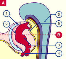
Fig. 2a - Sagittal section through an
embryo in stage 10 (roughly 28th day) |
|
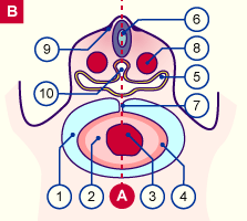
Fig. 2b - Transversal section through an
embryo in stage 10 (roughly 28th day) |
|
Legend |

1
2
3
4
5
|
Pericardial cavity
Cardiac jelly
Endocardal tube
Myoepicardium
Pharyngeal pouch |
|
|
|

6
7
8
9
10
|
Neural tube
Mesocardium
Aorta dorsalis
Neural crest
Notochord |
|
|
|
Fig. 2a
The cranial part of the embryo is shown in a medial sagittal section. The cardiac anlage is surrounded by the pericardial cavity. The heart consists of the myoepicardial mantle, the cardiac jelly and the endocardium.
Fig. 2b
In this transversal section the heart finds itself at the level of the rhomboencephalon, i.e., still very cranial. Note the position of the pericardium in relation to the heart, the many layers of the cardiac wall and the provisional existence of a dorsal suspension (mesocardium).
(see also the development of the pericardium)
|
|
More info
|
|
The outer layer of the heart is formed by the epicardium that originally arises from the extracardial anlage, the proepicardial serosa cells. ntrallyThese grow over the myocardium and issue from a collection of cells in the septum transversum that lie ventrally to the liver bud near the sinus venosus.
Presumably, the subepicardial mesenchymal cells also come from the epicardial layer (1).Besides the epicardial layer, it appears that the proepicardial serosal cells also form the endothelium and the smooth muscle cells of the coronary arteries. Moreover they play a modulating role in the development of the myocardium. (7)
|
|
|
|
|
|
|

