16.1 Outer form and position of the heart


The formation of the venticular loop - the U-shaped heart

Quiz

Quiz 12


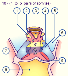
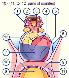
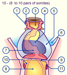
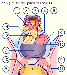
The early processes of cardiac formation, in particular the forming of the loop, are very interesting from many points of view. A long, closed tube arises out of the endocardial plexus. This tubular heart largely represents the anlage for the trabeculated part of the future right and left ventricles (3). The outflow tract, cone and the atrium make up here only a small part. Since the curvatures of the heart take place in various planes, the following interactive diagrams show them in two views.

Quiz

Quiz 25


The loop formation in a side view should mainly illustrate how the inflow tract (pink) slips behind the embryonic ventricle (violet) and the outflow tract (yellow). Further, the beginning formation of the sulcus atrio-ventricularis between the atrium and ventricle can be seen well.

Overview of the illustrations


In the frontal view mainly the formation of the D-loop of the embryonic ventricle (violet) is easily seen. The inflow tract moves upwards and will be covered by a part of the ventricle. The development of the sulcus primus can be easily recognized in this view. Via the movement of the inflow tract to the right a part (later: right atrium) becomes visible besides the outflow tract (Interactive diagram) .

Quiz

Quiz 04


Quiz

Quiz 05


Fig. 3 - Formation of the loop,
frontal view
Fig. 3 - Formation of the loop,
frontal view
 Legend


1
2
3
4
5
6
7
Pharyngeal pouch I
Aortic arch I
Foregut
Myoepicardial mantle
Pericardial cavity
Amniotic cavity
Endocardial loop


8
9
10
11
12
13
14

Midgut
Septum transversum
Omphalomesenteric vein
Umbilical vein
Sulcus primus
Sulcus atrio-ventricularis
later: right atrium

Fig. 3
Formation of the loop, frontal view of the embryonic heart


Video

The formation of the loop and the change in position of the individual cardiac segments that are connected with it can be followed by looking at the plastillin model.


Previous page | Next page