|

|
|
|
16.2 From serial to parallel circulation - the septation of the heart
|
|
|
|
|
|
The transformation processes of the atria
|
|
|
|
With the formation of the cardiac loop the inflow tract migrates with the sinus venosus and the two sinus horns up and to the rear. In addition, the vein opening that is originally located symmetrically is also shifted towards the right, together with its various branches.
In the beginning, the sinus venosus contains only blood from the two umbilical veins (oxygen- and nutrient-rich from the placenta), the omphalomesenteric veins and finally the commun cardinal veins, which receive the blood from the embryonic somatic circulation proper.
After various transformation processes in which the sinus venosus is incorporated into the right atrium and the left sinus horn atrophies, the blood gets directly into the right atrium through the superior and inferior vena cava.
The venous openings of the left sinus horn (common cardinal vein and umbilical / left omphalo-mesenteric vein) atrophy and the remnants form the sinus coronarius.
|
|
|
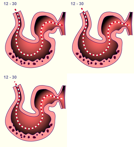
Fig. 10 - Side view
of a cardiac section |
|
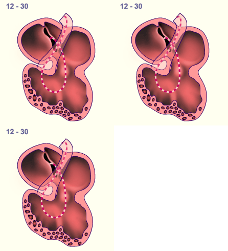
Fig. 11 - Ventral view
of a cardiac section |
|
Legend |

1
2
3
|
Ventricle
Atrium
Sinu-atrial inlet
|
|
|
|

4
5
6 |
Septum spurium
Dorsal septum atrio-ventriculare
in the atrio-ventricular canal
Sulcus atrio-ventricularis
|
|
|
|
Fig. 10, 11
In stage 12 (ca. the 30th day) the blood enters through the sinu-atrial inlet into the atrium and thereby gets into the common ventricle via the atrio-ventricular canal, which is outwardly constricted by the sulcus atrio-ventricularis
|
|
In this interactive diagram and using a front view and associated section, it is shown how the inflow tract is shifted to the right with the sinus venosus and the two sinus horns. The result is that the blood now only flows in the right part of the atrium (still common to both) through the sinu-atrial inlet. The blood flow through this opening is formed by two valves, the venous valves and their fusion forms the septum spurium above the opening.
|
|
|
In the heart itself various transformation processes are taking place at this time so that the blood no longer flows serially through it.
At the transition to the ventricle a prominent indentation named sulcus atrio-ventricularis forms between the atrium and ventricle segments. In the interior, the passage to the canalis atrio-ventricularis (av-canal) narrows. The shifting of the av-canal to the right has a decisive influence on the further development (it is now in the center and no longer on the left), so that the blood that initially only flowed in the left ventricle can now empty into both ventricles. Through this shift, a connection of the two atria to their ventricles has arisen. |
|
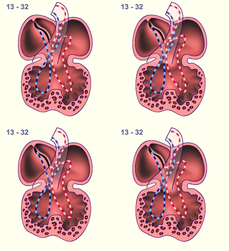
Fig. 12 - Ventral view of a cardiac section |
|
Legend |
|

1
2
3
4
5
6
7
8
|
Right atrium
Dorsal atrio-ventricular septum
in the av-canal
Right ventricle
Interventricular septum
Septum primum
Left atrium
Sulcus atrio-ventricularis
Left ventricle |
|
|
|
Fig. 12
In stage 13 (ca. the 32nd day) the av-canal is shifted in the middle of the heart and makes sure that each atrium is connected with its ventricle. |
In the atrium itself a fold, the septum primum  12-13 12-13 , grows to the left of the septum spurium from the roof of the still common atrium towards the av-canal. Only a small opening remains above the atrio-ventricular septum, the foramen primum. Due to the septum primum, the common atrium gets divided into right and left chambers. The common ventricle is also separated by the interventricular septum into two chambers. (see transformation processes of the ventricle). , grows to the left of the septum spurium from the roof of the still common atrium towards the av-canal. Only a small opening remains above the atrio-ventricular septum, the foramen primum. Due to the septum primum, the common atrium gets divided into right and left chambers. The common ventricle is also separated by the interventricular septum into two chambers. (see transformation processes of the ventricle). |
|
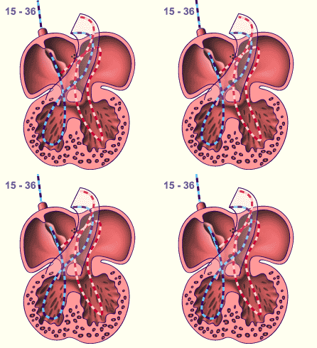
Fig. 13 - Ventral view of a cardiac section |
|
Legend |
|

1
2
3
4
5
6
7
8
9
10
11
12 |
Septum spurium
Sinu-atrial inlet
Dorsal atrio-ventricular septum
in the av-canal
Sulcus atrio-ventricularis
Right atrium
Interventricular foramen
Interventricular septum
Left ventricle
Right ventricle
Left atrium
Septum primum
Foramen secundum |
|
|
|
Fig. 13
The heart in stage 15 (ca. the 36th day) exhibits incompletely separated chambers.
The av-canal is divided by the dorsal (lower) and ventral (upper) cushion into left and right av-canals.
|
|
The flow relationships are such that the blood derived from embryonic somatic circulation, which flows into the right atrium via the common cardinal veins (later the superior vena cava), empties itself mainly via the right part of the av-canal into the right ventricle.
A further septum, the septum secundum, grows subsequently on the right of the septum primum from the front downwards and towards the lower rear. Through the newly created foramen secundum in the septum primum a connection still exists between the right and left atria. However, this connection becomes constricted and covered almost completely by the septum secundum until the end of the embryonic period. It is now called the foramen ovale and has a valve-like character in that the blood from the right side flows with a high pressure to the left side but no blood can return (more information in the interactive diagram).
|
|
|
Interactive diagram
|
|
In this interactive diagram the septation of the atria is shown in a sagittal section through the right
|
|
|
In summary it can be said that for the transformation from a serial to a parallel flow both outer as well as inner transformation processes are responsible. At the atrium level these are:
- The relocation of the sinus venosus to the right
- The relocation of the av level into the middle
- The septation of the common atrium
- The septum formation at the av level
|
|
|
|
|
|
More info
|
|
Today one can trace the origin of tissues using antibodies . With this method it can be shown that the tissue for the atrial septa comes from the left atrium. It appears that remaining mesenchymal cells that migrated via the mesocardium into the myocardium are decisive for the growth of the septa. One finds the corresponding gene expressing cells both there where the mesocardium atrophied as well as on the free edge of the septum primum. (8)
|
|
|
|
|
|
|

