|

|
|
|
16.2 From serial to parallel circulation - the septation of the heart
|
|
|
The septation of the outflow tract
|
|
|
|
In the course of further development of the heart the segment between the conus part and the aortic sac lengthens within the pericardium. Among other places, the material for this segment comes from neural crest cells that migrate over the pharyngeal arches III, IV and VI and are responsible for the development of the truncus arteriosus as well as the divisions of the outflow tract (9). Based on this fact, one always speaks in the following of the conotruncus.
|
|
|
| Fig. 16 - Migration of the neural crest cells via the pharyngeal arches |
|
Legend |
1
2
3
4
5
6
7
8
9 |
Neural crest
left internal carotid artery
First aortic arch in the first pharyngeal arch (mandibular arch)
Anlage of the eye
Conotruncus
Sixth aortic arch (pulmonary arch)
Aorta dorsalis
Aortic sac
Right ventricle |
|
|
|
Fig. 16
The neural crest cells migrate over the pharyngeal arches III, IV and VI along the aortic arches that have been formed to the heart. They are responsible for the lengthening of the truncus and the separation of the outflow tract.
|
|
More info
|
|
The pax3 gene is responsible for the fine-tuning of the neural crest cell emigration from the neural tube region. In the absence of the pax3 gene (splotch embryo) or by removal of the corresponding neural crest cells it has been shown experimentally that severe cardiac abnormalities result. (9)
|
|
|
|
|
|
The lengthening of the truncus part causes a further sulcus to occur between the conus and truncus that in the later development marks the formation of the endocardial cushions for the aorta and pulmonary valves (see semilunar valves). Very important for the division of the outflow tracts is the fact that the two streams of blood thus flow around each other spirally, influencing the growth of both the conus and truncus septa.
|
|
|
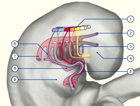
| Fig. 17 - Blood flow through the heart |
|
Navigation |
1
2
3
4
5
6
7
8
9
10
11 |
Aorto-pulmonary septum
Upper truncus septum
Right conus septum
Right av-canal
Rear processes of the interventricular septum
Aorta
Pulmonary truncus
Lower truncus septum
Left conus septum
Crista prima (almost atrophied)
Interventricular septum: anterior part |
|
|
|
Fig. 17
The right ventricle is cut open. One can see the not yet completely separated conotruncus.The spiral blood flow can be identified. |
|
Three components are responsible for forming the septa in the outflow tract (according to how the blood flows):
- Conus septum
- Truncus septum
- Aorto-pulmonary septum
For the conus and truncus septa initially only the endocardial cushions, formed through the influence of immigrated neural crest cells, are involved.They grow into the lumen and join up in the middle. The aorto-pulmonary septum consists of the mesenchyma between the pharyngeal arches IV and VI. (see also: development of the arteries).
It grows as an unpaired relief in a direction that is against that of the blood stream. Through the spiral-shaped blood flow its right process grows onto the lower and its left process towards the upper truncus septum and unites with it. The upper truncus septum grows against the flow onto the right conus septum and the lower towards the left conus septum.Thus the outflow tract septum has completed a rotation of 180 degrees.
Finally, the left conus septum joins with the free border of the septum interventriculare after the crista prima has regressed. The right conus septum joins with the dorsal av-septum
|
|
|
More info
|
|
In the interactive diagram the relationships of the outflow septation are shown in detail.
|
|
|
|
|