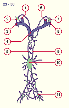
17.2 Lymphatic tissue


Lymph nodes


The formation of the lymph nodes (histological picture) follows that of the lymph vessels. The primary lymph nodes develop in regions that are occupied by lymphatic sacs. Between the branched lymphatic sacs mesenchymal solidifications form that join up incompletely. This leads to the formation of a plexus of lymph vessels. Fig. 12 - First lymphatic sacs in an
embryo of ca. 6 weeks
 Legend

1

2
3
4
5
6
7
8
Superior cardinal vein
(jugular vein)
Jugular lymphatic sac
Right subclavian vein
Axillary lymphatic sac
Left brachiocephalic vein
Thoracic duct (bilateral)
Lumbar lymphatic sac
Iliac lymphatic sac

Fig. 12
The development of the lymphatic system at 6 weeks. Three bilateral (jugular, axillary and lumbo-iliac) lymphatic sacs as well as the incompletely constructed lymph vessel plexus are shown.


As a first step - at the end of the embryonic period - the deep cervical lymph nodes form out of the jugular lymphatic sacs and soon afterwards the axillar, parasternal, mediastinal lymph nodes also form in the region of the shoulder girdle and the lymphatic nodes under the trapezius muscle.
The cysterna chyli, the retroperitoneal, lumbar and inguinal lymph nodes as well as the lymph nodes of the small pelvis develop out of the extended lumbar lymphatic sac.

Quiz

Quiz 07


Quiz

Quiz 11


Fig. 13 - Lymph plexus at the end of
the embryonic period (ca. 56 days)
Fig. 14 - Lymph plexus at the end of
the fetal period
 Legend

1
2

3
4
5
6
Right jugular vein
Right jugular and
axillary lymph duct
Right subclavian vein
Superior cava vein
Right thoracic duct
Left jugular vein


7

8
9
10
11
12
Left jugular and
axillary lymph duct
Left subclavian vein
Left thoracic duct
Cysterna chyli
Inguinal lymph nodes
Definitive thoracic duct

Fig. 13
The most important lymph vessels arise bilaterally and form a fine net.
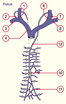
Fig. 14
Just as with the venous system the lymph vessels also atrophy unilaterally again.
In the breast-
abdominal space there remains only a thoracic duct at the junction of the jugular and subclavian veins that drains the lymph of the entire lower bodily region and the left head and arm region into the venous system. A smaller part, the lymph of the right-hand head-arm region, empties between the right jugular and subclavian veins into the venous system. The dashed lines indicate the atrophied parts of the lymph vessel system.


Soon, reticulum cells and macrophages also grow into these fetal lymph nodes and, somewhat later, the first lymphocytes appear as well as cells of the granulopoietic series. These lymph nodes also exhibit the first postcapillary venules.


Previous page | Next page