|

|
|
|
|
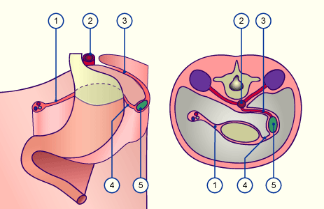
The spleen, a derivative of the mesoderm, is visible for the first time in stage 13 (ca. 32 days),  13 13 . It arises in direct contact to the stomach in the dorsal mesogastrium, which at this point is still very knobby. As the first differentiation arises a thickening of the visceral mesothelium and within it an accumulation of mesenchymal cells. Through the leftward shifting of the stomach the spleen is also moved to the left. The dorsal mesogastrium gets placed at the posterior abdominal wall and adheres in regions where the pancreas has grown in. Only the lieno-renal ligament remains, which connects the spleen with the posterior abdominal wall and contains the spleen vessels. The part of the original dorsal mesogastrium, which forms the connection between the spleen and the stomach, remains as the gastro-lienal ligament. Thus the spleen stays an intraperitoneal organ. . It arises in direct contact to the stomach in the dorsal mesogastrium, which at this point is still very knobby. As the first differentiation arises a thickening of the visceral mesothelium and within it an accumulation of mesenchymal cells. Through the leftward shifting of the stomach the spleen is also moved to the left. The dorsal mesogastrium gets placed at the posterior abdominal wall and adheres in regions where the pancreas has grown in. Only the lieno-renal ligament remains, which connects the spleen with the posterior abdominal wall and contains the spleen vessels. The part of the original dorsal mesogastrium, which forms the connection between the spleen and the stomach, remains as the gastro-lienal ligament. Thus the spleen stays an intraperitoneal organ.
|
|
|
| Fig. 15 - Formation of the spleen in the dorsal mesogastrium |
|
Legend |

1
2
3
4
5
|
Ventral mesogastrium
Aorta
Lieno-renal ligament
Gastro-lienal ligament
Spleen |
|
|
|
Fig. 15
The spleen is a mesodermal derivative and arises in the dorsal mesogastrium. Although parts of the dorsal mesogastrium adhere to the posterior abdominal wall, the spleen remains an intraperitoneal organ.
|
|
During the first trimenon additional types of cells migrate into the spleen, e.g., macrophages and precursor cells of the erythropoiesis. The mesenchymal cells begin to form a network. At this time hematopoiesis can also appear in the spleen. One also calls this developmental stage of the spleen a primary vessel reticulum. In the following weeks the spleen is modified and lobulated structures arise via the in-growth of trabeculae. After the 15th week the white spleen pulp with an accumulation of lymphocytes can be distinguished from the red spleen pulp that consists of the meshwork with sinusoids, described above, and lies in the periphery of the lobules.
|
|
|
|
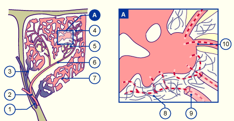
The colonization of the spleen with lymphocytes occurs at the beginning of the second half of the pregnancy. First T-lymphocytes surround the central arteries that diverge from the trabecular arteries. The further branchings of the blood vessels, the follicle arteries, lead to accumulations of B-lymphocytes. Additional, more in the periphery lying branchings, the penicillar arterioles and the sheathed capillaries bring blood via an open or closed circulation system into the peripheral sinusoids.
In the open circulation system, the capillaries discharge openly into the meshwork of a reticular fibrous framework (open circulation system). The blood must pass this meshwork before reaching the sinusoids. Thereby it is freed from old, no longer flexible erythrocytes. In a second circulation system the capillariess empty directly into the sinusoids. This represents the closed circulation system. The purified blood gets into the large circulation system again via the efferent veins.
|
|
|
| Fig. 16 - Spleen: scheme showing open and closed circulation |
|
Legend |

1
2
3
4
5
6
|
Trabecular veins
Trabecular arteries
Pulp veins
Penicillar arterioles
Follicle with follicle capillaries
Artery with periarterial lymphatic sheath (PALS) |
|
|
|
7
8
9
10
|
Splenic sinus
Reticular fibrous framework
Open capillaries
Closed capillaries |
|
|
|
|
|
Fig. 16
On the left diagram a section from a spleen is shown. One sees the branchings of the blood vessels.
On the right one sees a sinus with closed and open circulation systems.
|
|
|

