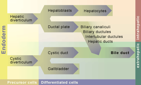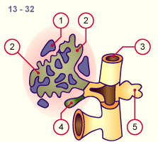
1
2
3
4
5
|
Capillary network of the
omphalomesenteric vein
Liver bud
Intestinal tube (duodenum)
Gall bladder
Dorsal pancreas anlage |
|
|
|
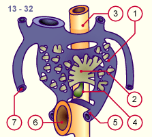
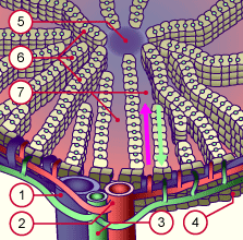
1
2
3
4
5
6
7
|
Capillary network of the
omphalomesenteric vein
Liver bud
Intestinal tube (duodenum)
Gall bladder
Omphalomesenteric vein
Omphalaomesenteric duct
Umbilical vein |
|
|
|
Fig. 32
Through sprouting and divisions of the intestinal bud, the first liver acini arise in the capillary network of the omphalo-
mesenteric vein.
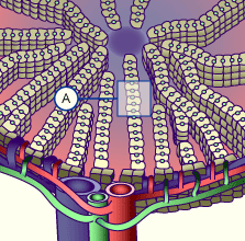
Fig. 33
Through the relocation of the entire cardiac circulation system to the right the liver also is shifted more into that position.
|