|

|
|
|
19.0 Learning aims, what you already should know, introduction, delving deeper
|
|
|
|
Students will be able to:
- describe the various parts that are involved in forming the face.
- trace the development of the teeth.
- explain the innervation of the tongue from an embryologic point of view.
- list the derivatives of the individual pharyngeal arches.
- construct the relationship between the aortic and pharyngeal arches.
- describe the individual portions of the intestine and know their definitive location in the abdomen.
- describe the mesenteric relationships with the associated intestinal sections and blood vessels.
- determine which blood vessel is responsible for which intestinal portion.
- map out the course of the portal vein and explain it from an embryologic point of view.
- know the individual parts of the pancreas and explain their derivation.
- draw the relationships of the duodenal loops in a fetus.
- discuss the development of the urogenital sinus with respect to the formation of the hind gut and anus
|
|
|
What you already should know
|
|
|
|
The development of the digestive tract begins with the formation of the definitive endoderm. In that the embryo folds laterally and bends the cranial and caudal ends ventrally, an intraembryonic tube arises from the endoderm that is distinct from the umbilical vesicle. This delimitation is explained by the enormous growth of the embryo, especially the neural tube.
One distinguishes three portions:
- Foregut
- upper section (embryonic pharynx)
- lower section (esophagus, stomach and upper part of the duodenum)
- Midgut
- Hindgut
The foregut is connected with the midgut via the anterior intestinal opening and the midgut to the hindgut via the posterior intestinal opening. In the midgut region the intestinal tube remains connected with the umbilical vesicle via the omphalomesenteric duct. The foregut is delimited by the prechordal plate at its cranial end. It dead-ends at a place that is only covered by ectoderm and this is termed the oropharyngeal membrane.
|
|
|
| Fig. 1 - Flexion of the embryo (stage 9, ca. 27 days) |
|
Legend |
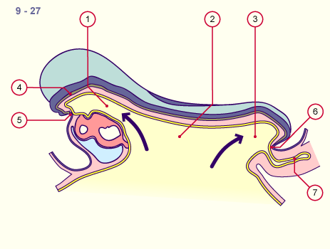
1
2
3
4
5
6
7
|
Foregut
Midgut
Rectum
Notochord
Oropharyngeal membrane / stomodeum
Cloacal membrane / proctodeum
Allantois |
|
|
|
Fig. 1
The embryo begins to fold itself ventrally. At this stage, the neural tube is not yet closed. The cardiac primordium, which initially lies in front of the embryo, has already turned 180 degrees.
|
|
With increasing formation of the tube through the embryonic flexion important new structures form in the foregut region due to the inductive influence of the mesenchyma around the intestinal tube. They arise through sprouting out of the endoderm. These are the thyroid, the thymus (laterally out of pharyngeal pouches 3 and 4) and the lungs in the upper section of the foregut (pharynx).
In the lower part of the foregut (esophagus, stomach and the cranial part of the duodenum), the liver and the pancreas (both shown in Fig. 3) develop.
|
|
|
| Fig. 2 - Flexion of the embryo (stage 10, ca. 28 days) |
|
Legend |
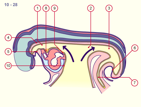
1
2
3
4
5
6
7
8
9
10 |
Foregut
Midgut
Rectum
Notochord
Oropharyngeal membrane / stomodeum
Cloacal membrane / proctodeum
Allantois
Primordium (or anlage) of the thyroid
Primordium (or anlage) of the lungs
Primordium (or anlage) of the liver |
|
|
|
Fig. 2
Around the cardiac primordium various sproutings occur out of the endoderm of the intestinal anlage. The transition from the intraembryonic to the extraembyonic parts of the endoderm becomes more and more constricted and will form a portion of the umbilical cord.
|
|
The hindgut also dead-ends in the caudal bud and there borders directly on the ectoderm, at a place where no mesoderm lies in between. This location is termed the cloacal membrane. With the flexion the allantois is also pushed in a ventral direction and gets taken into the embryonic body. The caudal end of the hindgut enlarges to become the cloaca, into which the allantois also discharges.
|
|
|
| Fig. 3 - Flexion of the embryo (stage 13, ca. 32 days) |
|
Legend |
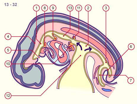
1
2
3
4
5
6
7
8
9
10
11
12
13 |
Foregut
Midgut
Rectum
Notochord
Oropharyngeal membrane / stomodeum
Cloacal membrane / proctodeum
Allantois
Primordium (or anlage) of the thyroid
Primordium (or anlage) of the lungs
Primordium (or anlage) of the liver
Primordium (or anlage) of the dorsal pancreas
Omphalomesenteric duct
Primordium (or anlage) of the stomach |
|
|
|
Fig. 3
The omphalomesenteric duct as well as various anlagen for endodermal derivatives have arisen. The anterior intestinal openings are present while the posterior one is still a dead end.
|
- How, in the area of the face, can epithelial bridges partly form again that make isolated facial folds disappear?
- How is it that the ocular primordia (or anlagen) and nasal placodes are brought to the middle?
- How do isolated primordia (or anlagen) migrate caudally from out of the pharyngeal pouches while others remain in the neck region?
- What causes the stomach to form?
- How does it happen that the intestines are turned towards the right?
- What influences cause individual portions of the intestines to develop differently?
|
|
|
|
|