|

|
|
|
|
The midgut extends from the apex of the duodenal loop, which is fixed to the large liver anlage via the bile duct, to the last third of the transverse colon.
Its parts are:
- Inferior part of the duodenum with the duodeno-jejunal bend
- Jejunum
- Ileum with the iliocaecal valve
- Cecum with vermiform appendix
- Ascending colon
- Transverse colon (2/3)
The midgut is supplied with blood by the superior mesenteric artery and innervated by the vagus nerve (CN X). Within the whole midgut and rectum unit there exists only one dorsal mesenterium, the ventral being readsorbed. Differentiation occurs in a cranio caudal sequence within a time window of roughly one week.
|
|
|
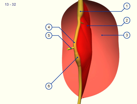
| Fig. 19 - Intestinal rotation: stage 13, ca. 32 days |
|
Legend |
1
2
3
|
Stomach
Mesenterium
Parietal peritoneum |
|
|
4
5
6 |
Intestinal loop
Omphalomesenteric duct
Cecum |
|
|
|
|
Fig. 19
The intestinal tube becomes enwrapped by the visceral peritoneum that connects it to the posterior body wall forming the dorsal mesenterium (red surface).
In this stage the intestinal tube is almost straight and is connected to the umbilical vesicle by the omphalo-
mesenteric duct.
|
|
|
|
|
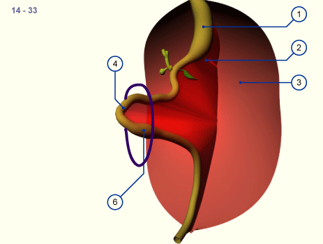
Only when the umbilical loop lengthens and grows into the umbilical coelom does it experience a rotation of 90 degrees in a clockwise direction as seen from the embryo. The cranial pedicle comes to lie to the right and the caudal to the left (stage 14, ca. 33 days,  14 14 ). The umbilical loop now has a horizontal position. Through the cranio-caudal growth gradient, the cranial pedicle forms first through lengthening of several loops in the umbilical coelom. ). The umbilical loop now has a horizontal position. Through the cranio-caudal growth gradient, the cranial pedicle forms first through lengthening of several loops in the umbilical coelom.
|
|
|
| Fig. 20 - Intestinal rotation: stage 14, ca. 33 days |
|
Legend |
1
2
3
|
Stomach
Mesenterium
Parietal peritoneum |
|
|
|
|
|
|
Fig. 20
The navel opening is schematically indicated by the blue ring. The developing intestines invade the abdominal space, gliding into it.
|
|
|
|
|
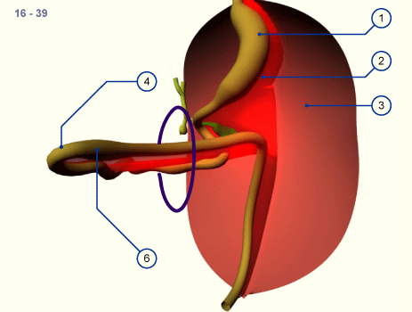
The developing umbilical loop extends further into the umbilical coelom because there is no more room for it within the embryo's abdominal cavity. It is the time of the strongest flexion of the embryo. Very soon a thickening in the region of the caudal pedicle of the intestinal tube is also to be seen: the cecum. Visually, it becomes an important fixed point for purposes of orientation.
|
|
|
| Fig. 21 - Intestinal rotation: stage 16, ca. 39 days |
|
Legend |
1
2
3
|
Stomach
Mesenterium
Parietal peritoneum |
|
|
|
|
|
|
Fig. 21
The entire intestinal loop has relocated in the umbilical coelom due to the limited space conditions in the abdominal cavity. The intestinal loop now has a horizontal orientation and the lengthening tube has formed several loops in the cranial pedicle. The caudal part is still straight.
|
|
|
|
|
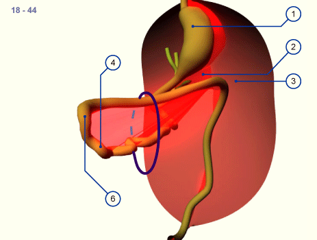
As development proceeds the intestinal loop turns further around its own axis. In stage 18 (ca. 44 days,  18 18 ) the extension of the intestinal loop into the umbilical coelom has reached its maximum. This physiologic navel hernia remains in existence up to the 9th week of pregnancy. (Omphalocele / umbilical hernia) ) the extension of the intestinal loop into the umbilical coelom has reached its maximum. This physiologic navel hernia remains in existence up to the 9th week of pregnancy. (Omphalocele / umbilical hernia)
|
|
|
| Fig. 22 - Intestinal rotation: stage 18, ca. 44 days |
|
Legend |
1
2
3
|
Stomach
Mesenterium
Parietal peritoneum |
|
|
|
|
|
|
Fig. 22
The largest part of the intestinal loop lies in the umbilical coelom and several loops have formed through the lengthening in the cranial, small intestine region.
|
|
|
|
|
At first, the loops of the small intestine return into the abdominal cavity and come to lie in the left half surrounded by the horizontal and descending part of the colon that never left the abdominal cavity. The rotation now amounts to more than 180 degrees and the colon is also shifted more and more into the abdominal space. The repositioning of the physiologic umbilical hernia is facilitated by the righting of the embryo's body.
|
|
|
| Fig. 23 - Intestinal rotation: stage 20, ca. 49 days |
|
Legend |
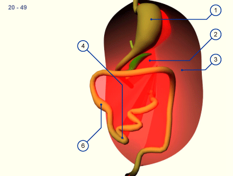
1
2
3
|
Stomach
Mesenterium
Parietal peritoneum |
|
|
|
|
|
|
Fig. 23
With the return of the intestines into the abdominal cavity the small intestine is moved to the left side and the cecum and the ascending part of the large intestine to the right. Initially the cecum may possibly be found in the upper right quadrant (elevated cecum).
|
|
|
|
|
Thus, after the reintegration of the intestinal loops into the abdominal cavity from the physiologic umbilical hernia, the derivatives of the originally caudal pedicle occupies the upper and ventral part of the abdominal cavity. At the end of the embryonic period this part migrates downwards into the iliac fossa, whereby an additional rotation occurs. The whole rotation of the intestines thus amounts to approximately 270 degrees. As a consequence, the mesenterium also turns with it and in its insertion it crosses over the inferior part of the duodenum. (Malrotation and congenital high cecum)
|
|
|
| Fig. 24 - Intestinal rotation: stage 23, ca. 56 days |
|
Legend |
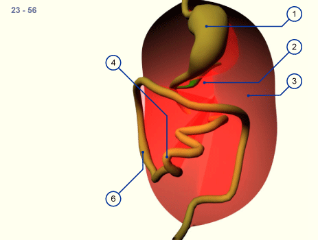
1
2
3
|
Stomach
Mesenterium
Parietal peritoneum |
|
|
|
|
|
|
Fig. 24
As a rule the cecum grows caudally and comes to lie in the right iliac fossa. Through rotation of the whole small intestine of more than 270 degrees the mesenterium also rotates thereby and moves off from the posterior wall over the inferior part of the duodenum to the small intestine.
|
|
|
|
|
|

