|

|
|
|
20.4 Lower urinary system
|
|
|
|
We have seen that the upper urinary system - consisting of the collecting ducts, the calices, the renal pelvis, and the ureters - arises from the ureter anlage.
The lower urinary system - composed of the bladder and the urethra - is formed from the endoderm of the posterior intestine.
|
|
|
Fig. 19 - Development of the cloaca,
Stage 13, roughly 32 days |
|
Fig. 20 - Migration of the kidneys,
Stage 23, roughly 56 days |
|
Legend |
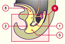
1
2
5
8 |
Urorectal septum
Cloacal membrane
Cloaca
Allantoïs |
|
|
|
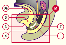
3
4
6
7
8a
|
Urogenital orifice
Anal orifice
Urogenital sinus
Rectum
Future bladder |
|
|
|
Fig. 19
The white arrow indicates the direction of growth of the urorectal septum. This separates the cloaca into a urogenital sinus (ventrally) and the rectum (dorsally).
Fig. 20
In this diagram the kidneys are found in their definitive position at the level of the upper lumbar region. The urorectal septum (white arrow) has divided the cloaca. The urogenital sinus, bounded on the outside by the urogenital orifice, lies ventrally. The rectum, which opens to the outside through the anal orifice, is found behind it. The cloacal membrane disappears in stage 19.
|
|
The perineum and the urorectal septum
|
|
|
|
Today, the urorectal septum is no longer regarded as an isolated cellular layer of mesoderm cells that slowly grow towards the cloacal membrane. It consists of two mesodermal structures that are fused together.
An upper fold (Tourneux), located frontally, grows caudally. Near the cloacal membrane two lateral folds (Rathke) form that fuse at the median level.
|
|
|
| They subdivide the cloaca and also grow in the direction of the upper fold (Tourneux) that is located frontally. A disorder in the formation of these two structures leads to recto-urethral or rectovesical fistulas. Connective tissue and the perineal musculature, which keep the pelvic organs in place, arise from the mesoderm, which surrounds the rectal tube. The central fibrous part of the perineum corresponds anatomically to the region between the anal and urogenital orifices. |
|
Fig. 21 - Development of the urorectal septum |
|
Legend |
|
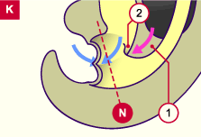
1
2
|
Peritoneal cavity
Upper fold (Tourneux) (pink arrow)
Lower fold (Rathke) (blue arrow) |
|
|
|
Fig. 21
The urorectal septum consists of two different mesoderm structures.
Over the course of the 4th week an upper fold (Tourneux) divides the cloaca in the cranio-caudal direction. The two lower, lateral folds (Rathke) are responsible for the division in the lower section.
|
Fig. 22 - Separation of the cloaca
and the formation of the perineum |
|
Fig. 23 - Separation of the cloaca
and the formation of the perineum |
|
Legend |
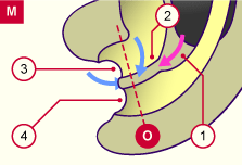
1
2
3
4
|
Peritoneal cavity
Upper fold (Tourneux; pink arrow) ; Lower folds (Rathke; blue arrows)
Primary urogenital sinus
Anal canal |
|
|
|
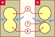
5
6
7 |
Urogenital orifice
Anal orifice
Perineum |
|
|
|
Fig. 22
When the two lower folds (Rathke) are fused in the median level, cranially meet at the upper fold (Tourneux), and merge with it, the separation of the cloaca is completed. (see the transverse section in Fig. 23 O).
Fig. 23
Transverse section from Figs. 21 and 22 at the N and O levels.
|
|
|

