19.4 Hindgut


Introduction

Quiz

Quiz 14


The hindgut extends from the left third of the transverse colon to the cloaca (rectum). In contrast to the midgut, no intestinal rotation occurs here but rather this part gets pushed to the left side by the midgut returning from the umbilical coelom. It consists of:

  • Transverse colon (left third)
  • Descending colon
  • Sigmoid colon
  • Rectum
  • Anal canal


Hindgut


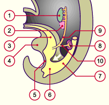
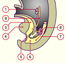
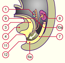
Initially the hindgut dead-ends in the cloaca and is separated by the cloacal membrane from the ectodermal anal pit, the proctodaeum. In addition, at its end, it is connected to the allantois and to the mesonephric duct.
From stage 14, ca. 33 days, 14 the cloaca flattens in the frontal plane and extends somewhat in the sagittal plane, whereby from the upper rear and from both sides a mesenchymal condensation, the urorectal septum, arises in the angle between the allantois and the hindgut. Through this mechanism, the cloaca is subdivided into the urogenital sinus (ventrally) and the anorectal canal (dorsally) (2). (See details about the subdivision of the urorectal septum in the urinary system module).


Fig. 25 - Hindgut in stage 15
(ca. 36 days)
Fig. 26 - Hindgut in stage 18
(ca. 44 days)
 Legend

1
2
3
4
5
6
7
8
9
10
Gonads
Mesonephros
Allantois
Genital tubercle
Cloacal membrane
Cloaca
Hindgut
Metanephros
Mesonephric duct (Wolff's duct)
Ureter


1
3
4
5
6
7
8
9
Gonads
Anlage of the bladder
Genital tubercle
Cloacal membrane
Rectum
Hindgut
Metanephros
Mesonephric duct (Wolff's duct)

Fig. 25
The urorectal septum extends itself caudally (in the direction of the arrow) and subdivides the urorectal sinus into a urogenital sinus and an anorectal canal.

Fig. 26
The urorectal septum has almost reached the perineum.


In the cloacal region the urorectal septum thickens still more and forms the material for the deep perineum. In stage 19, ca. 46 days, 19 the cloacal membrane ruptures and the endodermal part of the anal canal goes over into the ectodermal part of the proctodeum. During this period the anorectal canal is closed by an epithelial plug and is recanalized again only in the early fetal period. Fig. 27 - Hindgut in stage 23
(ca. 56 days)
 Legend

1
3
4
6a
7
8
9
10a
11
12
Anlage of the gonads
Anlage of the bladder
Genital tubercle
Rectum
Hindgut
Metanephros
Mesonephric duc (Wolff's duct)
Ureter
Urogenital orifice
Anal orifice

Fig. 27
The separation is now complete and the cloacal membrane ruptures in this stage.


More info

The first villi and Lieberkühn's glands arise in the early fetal period. However, they disappear shortly before birth and appear again only after birth. With the beginning of the second trimester the meconium in the large intestine is formed. It consists of dark green material that mainly consists of a mixture of secretions of the intestinal glands and the gall bladder as well as of amniotic fluid, which is swallowed by the fetus. The intestinal content is sterile up to birth. Following birth the meconium is passed as the first stool by the newborn.


List of the chapters | Next chapter