|

|
|
|
21.3 Differentiation of the gonads
|
|
|
|
The differentiation of the ovaries happens later than that of the testes, taking place during the 8th week  20-23 20-23 . Since females lack the Y chromosome, they have no SRY gene, except when a translocation of the gene onto the X chromosome occurs! . Since females lack the Y chromosome, they have no SRY gene, except when a translocation of the gene onto the X chromosome occurs!
Histologically two regions can be distinguished in an ovary:
- Cortex, containing all the elements of the parenchyma
- Medulla, which shares the elements of the stroma with the cortex.
|
|
|
More info
|
|
Special case of a dissociation between a phenotype and a genotype
|
|
|
|
Development of the stroma
|
|
|
|
In an ovary the majority of the gonadal cords stay in contact with the surface coelomic epithelium. Those gonadal cords that go into the depths out of the thickened coelomic epithelium and lose contact with it atrophy. One also suspects there are signals from the ovary, though, which actively prevent the differentiation into male gonads. So, for example, WNT-4 functions partly as an anti-testis gene in that it suppresses certain developmental steps of differentiation in the direction of the testes (16).
|
|
|
Fig. 15 - Indifferent gonads
stage 17, ca. 41 days |
|
Fig. 16 - Early differentiation (female)
stage 20, ca. 49 days |
|
Legend |
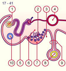
1
2
3
4
5
6
7
8
9
10 |
Mesonephric duct (Wolff)
PGC
Peritoneal cavity
Aorta
Mesonephric tubule
Local coelom mesenchyma
Thickened coelomic epithelium
Intestine
Mesentery
Anlage of the paramesonephric
duct (Müller) |
|
|
|
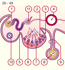
1
2
3
4
5
6
7
8
9
10
11 |
Mesonephric duct (Wolff)
PGC
Peritoneal cavity
Aorta
Mesonephric tubule
Degenerated gonadal cords
Thickened coelomic epithelium
Intestine
Mesentery
Anlage of the paramesonephric
duct (Müller)
Atrophy of the mesonephric
nephron |
|
|
|
Fig. 15
The PGC immigrate into the genital ridge. Gonadal cords continue to develop and become infiltrated with PGC. At this time, the mesonephric
structures develop into the mesonephric ridge. The paramesonephric duct is also formed.
Fig. 16
Those gonadal cords, which extend into the depths, atrophy. Only the gonadal cords, which stay in contact with the coelomic epithelium, remain.
|
|
Development of the parenchyma
|
|
|
|
Towards the end of the embryonic period one can distinguish the cortex with its gonadal cords and the medullar PGC in the ovary. Although one often finds in the literature that cells with a mesonephric origin are also present in the ovarian stroma, examinations made on mice ovaries have shown that no cells migrate out of the mesonephros into the ovary (in contrast with myofibroblasts of the testis) (17). Probably the cells of the mesonephros only reach the hilus area of the ovary and participate there in the weakly formed rete ovarii.
In the course of the 4th month the gonadal cords dissolve - also in the cortex - due to blood vessels that are sprouting from the medulla and isolated cell accumulations surround the oogonia that increasingly divide synchronously (mitosis). Like the spermatogonia the oogonia form similar cell clones. The individual cells are connected with each other via cellular bridges. One can now distinguish various zones in the cortex: In the outermost zone proliferating oogonia are found; somewhat further inward one recognizes oocytes that have spontaneously entered into the prophase of the first meiosis (meiosis 1).
From the 5th month a third zone becomes visible towards the medulla in which the oocytes have already completed the prophase of the first meiosis and are surrounded by a monolayer of cells that have differentiated out of the gonadal cord cells and now are now called follicle or granulosa cells. The primary oocytes that are enveloped by follicle cells are now designated primordial follicles and then remain in this stage of the first meiosis (dictyotene stage).
|
|
|
Fig. 17 - Early differentiation (female)
stage 23, ca. 56 days |
|
Fig. 18 - Further differentiation (female)
ca. 20 weeks |
|
Legend |
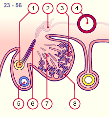
1
2
3
4
5
6
7
8
|
Mesonephric duct (Wolff)
Mesonephric nephron atrophying
Oogonia in the ovarian cortex
Aorta
Paramesonephric duct (Müller)
Mesonephric tubules atrophying
Degenerated gonadal cords
Thickened coelomic epithelium in
contact with the gonadal cords |
|
|
|
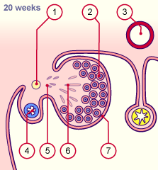
1
2
3
4
5
6
7 |
Mesonephric duct (Wolff)
atrophying
Primordial follicle in the ovarian
cortex
Aorta
Paramesonephric duct (Müller)
Mesonephric tubules atrophying
Degenerated gonadal cords
Mesothelium of the ovary |
|
|
|
Fig. 17
The gonadal cords in the center of the ovary degenerate and only those that are near the surface epithelium remain. Through blood vessels that grow into it and connective tissue stroma from the medulla the gonadal cords in the cortex break up into small cell accumulations. These increasingly surround the PGC and the primary oocytes that also further develop in the cortex.
Fig. 18
After approximately the middle of the pregnancy (20th week) the entire cortex is filled uniformly with primordial follicles. The rete ovarii receives no connection to the mesonephric tubules or the mesonephric duct. |
|
|
|
|
Comparison table
|
|
Comparison table of the differentiation of the gonads in males and females.
|
|
|
|
|
|
During the early fetal period millions of primordial follicles arise through intensive mitotic divisions of the oogonia (the follicle stages from primordial follicle to tertiary follicle).
The number of the primordial follicles at birth amount to between 300,000 and 2 million, but they decrease massively from then till puberty. At the beginning of puberty only ca. 40,000 still remain. Of these only ca. 300 primary oocytes develop further between puberty and menopause into fertilizable oocytes.
It is to be noted that follicles only form in the presence of the PGC. Without them, sterile gonadal cords are formed that then further degenerate and as a consequence the ovary then consists only of stroma.
|
|
|
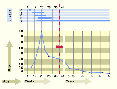
| Fig. 19 - Development of the primordial germ cells (PGC) |
|
Legend |
|
|
Fig. 19
The upper part of the graph shows the approximate duration of the various phases of PGC development.
The lower part shows the development of the number of PGC and primary oocytes.
|
|
Phase A:
A migration of the PGC into the genital ridge where they mitotically multiply through contact with the coelomic epithelium. Formation of the gonadal cords that partially degenerate (weeks 6-7).
Phase B:
Active proliferation phase of the PGC and differentiation into oogonia (weeks 9 – 22). The maximal number of PGC (7 million) is attained with 20 weeks.
Phase C:
The oogonia enter spontaneously into meiosis and become arrested in the diplotene of the prophase of the first meiosis. One now designates them as primary oocytes (weeks 12-25).
Phase D:
Formation of primordial follicles (weeks 16 -29)
Phase E:
Progressing follicular atresia from the 16th week on. Between 300,000 and 2 million primordial follicles remain up to birth and of these ca. 300 primordial follicles develop further between puberty and menopause, in order to let fertilizable oocytes arise. The majority of the follicles thus perish along the way.
|
|
|
The development of the ovary is characterized by the following:
- Gonadal cords remain in existence only near the cortex; in the medulla they atrophy. The rete ovarii is only rudimentarily developed. Its cells probably come from the mesonephros, but a connection between the rete testis and the mesonephros never comes into existence.
- Cortical gonadal cords separate themselves from the others in order to make isolated cell accumulations around the oocytes and thus to form the primordial follicles.
- Coelomic epithelium, out of which a simple cubic ovarian mesothelium arises.
|
|
|
|
|
|
|