|

|
|
|
21.3 Differentiation of the gonads
|
|
|
|
The testes differentiate themselves earlier than the ovaries, namely in the course of the 7th week  18 18 (44 days). (44 days).
Responsible for this is the SRY gene on the Y chromosome that induces the development of the testes through the activation of a series of further genes (sex-determining genetic factors and hormones).
|
|
|
|
Development of the parenchyma
|
|
|
|
The differentiation of Sertoli's supporting cells form the first step in the organogenesis of the testes. These cells come - in any case in mice (14) - from pluripotent coelomic epithelial cells of the gonadal ridge. In the gonadal anlage, through the influence of genetic products that are activated by the SRY, they form intercellular membrane connections and in this way surround more and more the primordial germ cells, while growing at the same time as gonadal cords into the medulla. In addition, in a male embryo, cells of mesonephric origin are involved as well in forming the gonadal cords, by accumulating on the outside of the gonadal cords and forming the peritubular myoblasts. From the gonadal cords the testicular cords form that then differentiate to become the convoluted seminiferous tubules (500 to 1000) and straight seminiferous tubules of the mature testicles.
|
|
|
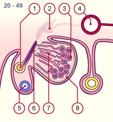
Fig. 10 - Early differentiation (male)
stage 18, ca. 7 weeks |
|
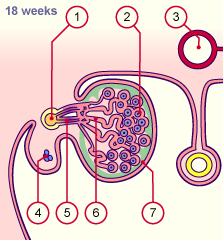
Fig. 11 - Early differentiation (male)
stage 20, ca. 8 weeks |
|
Legend |

1
2
3
4
5
6
7
8
9
10
11 |
Mesonephric duct (Wolff)
PGC
Peritoneal cavity
Aorta
Mesonephric tubule
Gonadal cords
Coelomic epithelium
Intestine
Mesentery
Anlage of the paramesonephric
duct (Müller)
Mesonephric nephron |
|
|
|
1
2
3
4
5
6
7
8
|
Mesonephric duct (Wolff)
Mesonephric nephron
(atrophying)
Testicular cords surround the PGC
Aorta
Paramesonephric duct (Müller)
Mesonephric tubule
Testicular cords that grow into the
medulla
Tunica albuginea |
|
|
|
Fig. 10
The gonadal cords (future testicular cords) increase in the depths while the nephrons of the mesonephros slowly atrophy. Only the distal part of the mesonephric tubules stays in existence in the male gonads.
Fig. 11
In the periphery the testicular cords surround the PGC and, in the depths, take up contact with the mesonephric tubules. Between the testicular cords and the surface epithelium the tunica albuginea, a connective tissue capsule that envelops the testes, forms.
|
Fig. 12 - Late differentiation (male)
ca. 18 weeks |
|
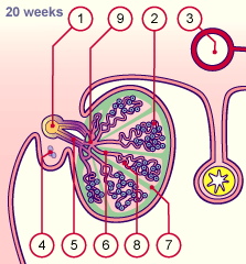
Fig. 13 - Late differentiation (male)
ca. 20 weeks |
|
Legend |
1
2
3
4
5
6
7
|
Mesonephric duct (Wolff)
Testicular cords, surround the PGC
Aorta
Paramesonephric duct (Müller)
(atrophying)
Mesonephric tubule
(later efferent ductules)
Testicular cords
Tunica albuginea |
|
|
|
1
2
3
4
5
6
7
8
9 |
Mesonephric duct (Wolff)
PGC surrounded by supporting
cells (Sertoli)
Aorta
Paramesonephric duct (derivative)
Efferent ductules
Straight seminiferous tubule
Tunica albuginea
Convoluted seminiferous tubule
Rete testis (testicular network) |
|
|
|
Fig. 12
The testicular cords penetrate into the medulla, branch within the tunica albuginea, and form anastomoses among themselves and with the mesonephric tubules, leading to the formation of the rete testis.
Fig. 13
The deep portion of the testicular cords form the straight seminiferous tubules, which converge to the rete testis, from which - on the other side - the efferent ductules (mesonephric tubules) depart. Finally, they empty into the mesonephric duct (Wolff).
|
|
Until puberty the coiled testicular cords are filled. During puberty they form lumens and are from then on called convoluted seminiferous tubules. The germ cells on the other hand divide mitotically, but their meiosis begins only with puberty. The deep portions of the coiled testicular cords, which are delimited by septa, are stretched and are called straight seminiferous tubules. These last go over into the rete testis, which is a labyrinth of small passages in the tunica albuginea. The thin wall possesses a cubic epithelium. During the 9th week, from 5-12 mesonephric tubules the efferent ductules form that bind with the rete testis in the 3rd month.
|
|
|
Overview
|
|
Comparison of the differentiation of the gonads between male and female.
|
|
|
| Fig. 14 - Detailed diagram of the differentiated testis in the 4th month |
|
Legend |
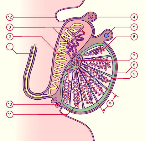
1
2
3
4
5
6
7 |
Deferent duct (Wolff)
Epididymis
Efferent ductules
Appendix epididymidis
Appendix testis
Convoluted seminiferous tubules
Rete testis |
|
|
|
8
9
10
11
12
A
|
Straight seminiferous tubules
Tunica albuginea
Paradidymis
Interlobular septum
Mesothelium
Lobule |
|
|
|
|
|
Fig. 14
The deep medullary portion of the testicular cords are stretched and converge towards the rete testis, which on the other side goes over into the efferent ductules (mesonephric tubules) that go out from the mesonephric duct (Wolff). From the 8th week the latter form compact convolutions, the ductus epididymidis. Outside the epididymis this continues as the deferent duct. The tunica albuginea is now a taut connective tissue layer that envelops the testicles. Connective tissue septa subdivide the testicles into lobules.
In this illustration, one also sees the embryonic derivatives of the paramesonephric duct (appendix testis), the mesonephric duct (appendix epididymidis) and the mesonephric tubules (paradidymis).
|
|
The efferent ductules form the connection between the rete testis and mesonephric duct. Towards the end of the 8th week, under the influence of testosterone, the cranial part of the mesonephric duct gets to be tightly coiled and so forms the ductus epididymidis which, outside the epididymis, continues as the deferent duct. (see: the inner sex passages)
|
|
|
|
After the 8th week certain mesenchymal cells between the testicular cords differentiate to become interstitial cells (Leydig), which produce testosterone. The testes thus represent an endocrine gland that produces androgens. The origin of these cells is still unclear – one suspects that a steroid-producing population of cells in the ventral part of the mesonephros differentiate and form both the origin of the adrenal cortex cells and also interstitial cells (Leydig) (15).
|
|
|
|
Development of the stroma
|
|
|
|
The mesenchyma between the testicular cords congeals and forms connective tissue septa that subdivide the testicles into lobules (ca. 250-370).
In stage 22 22 (ca. 53 days) this mesenchyma also forms a taut connective tissue layer between the testicular cords and the coelomic epithelium as well as the future tunica albuginea. Finally, the coelomic epithelium transforms itself into a mesothelium, just like the coelomic epithelium around the other serous cavities (peritoneum, pleura, pericardium).
|
|
|
Summary:
- In the testicular cords PGC (future spermatozoa) are to be found. The somatic cells differentiate themselves into Sertoli's supporting cells, responsible for nourishing the spermatozoa and secreting the antimüllerian hormone (AMH), which promotes the atrophy of the paramesonephric duct (Müller).
- The rete testis forms the continuation of the centrally-lying testicular cords or the straight seminiferous tubules.
- The efferent tubules connect the rete testis with the mesonephric duct (Wolff), the future epididymis, which continues with the deferent duct.
- The interstitial mesenchymal cells of the testes develop into Leydig's interstitial cells. They are responsible for the production of testosterone that, among other things, assures that the mesonephric duct (Wolff) does not atrophy.
- The stroma, made of connective tissue, subdivides the testes into lobules and forms the tunica albuginea.
|
|
|
|
|
|
|