21.4 Development of the internal genital organs


In males: Differentiation of the canal system in the genital organ

Quiz

Quiz 11


The differentiation of the male sex organ canals is influenced by the testosterone hormone that is produced from the 8th week in the fetal testis by Leydig's interstitial cells.


Fig. 36 - Differentiated canal system of the male sex organs, ca. 3rd month  Legend

1
2
3
4
5
6
7
8
9
10
A
Epididymal appendage
Testicular appendage
Atrophying paramesonephric duct (Müller)
Deferent duct
Seminal vesicle
Ejaculatory duct
Paradidymis
Gubernaculum
Prostatic utricule
View of the dorsal side of the urogenital sinus (=> prostatic utricule)
Seminal colliculus

Fig. 36
The paramesonephric duct (Müller) atrophies (shown in blue).
The mesonephric duct (Wolff) differentiates itself further (shown in pink).
The two ejaculatory ducts as well as the prostatic utricle (which will have a dead end due to the atrophy of the paramesonephric duct) empty into the seminal colliculus.


Two phenomena mark the differentiation of the canals of the internal male sex organs:

  1. The atrophy of the paramesonephric duct (Müller)
  2. The development and differentiation of the mesonephric duct (Wolff)

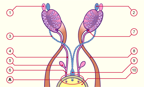
The mesonephric duct (Wolff) atrophies cranially and leaves behind only the epididymal appendage as an embryonic rudiment. On both sides the parts of the mesonephric duct, which lie across from the testes, form the epididymis. The testis and the epididymis of both sides are partially enveloped by the tunica vaginalis testis (serous bilaminar membrane with a periorchium [ = outermost layer] and epiorchium [ = inner layer]). In the part of the epididymis are end the efferent ductules. They originate from the mesonephric tubules, and so form the beginning of the epididym. Immediately afterwards it coils tightly and finally goes into the lower part of the epididymis (its tail) and over into the deferent duct. This is a musculo-epithelial tube that, during ejaculation, dispatches the sperm cells from the epididymis into the urethra.

Histology

Deferent duct


In the male fetus - between the 8th and 11th week - the paramesonephric duct atrophies due to the effects of the antimüllerian hormone (AMH), which is formed by precursors of Sertol's supporting cells (cells that surround the primordial germ cells and come from the primitive gonadal cords). Despite the effects of this hormone, embryonic remnants of the paramesonephric duct remain behind in males. These are the testicular appendage at the cranial pole of the testis and the prostatic utricle at its caudal pole.


Overview table

Overview table comparing the differentiation of the internal genital organ canals of both sexes.


Previous page | Next page