|

|
|
|
|
The abnormalities of the sexual organs are manifold.
They can be caused by:
- Environmental factors (the incidence of cyptorchism has doubled in the last 30-50 years and that of malignant tumors of the testes even more than doubled) (8, 9)
- Genetic disorders (absent disjunction of the sex chromosomes)
- Hormonal factors
|
|
|
Pathology of sexual differentiation conditioned by genetic or hormonal causes
|
|
|
|
When the development in the one or other sex is interrupted in the embryonic stage a hermaphroditism develops. Thereby, histologically, one finds ambiguous gonads and the external genitalia are also ambiguously defined.
|
|
|
|
True hermaphroditism is extremely rare. An individual is involved who simultaneously possesses both testicular and ovarian tissues.
Ethiology:
The karyotype of hermaphrodites exhibits an alteration of the gonosomes. Thereby mosaics (ca. 30%) of two cell populations (with XX and XY) are found, which probably is to be traced back to a fusion of two zygotes of different sexes. 60% of the patients have a 46,XX karyotype, while 10% exhibit a 46,XY karyotype. With the 46,XX karyotype molecular investigations have shown that in the fewest cases a SRY gene can be detected. This leads one to suspect that the disorder is probably connected with an X-bound mutation that is responsible for the development of the testes.
Various mechanisms are imaginable for this pathology, but at this time one knows still relatively little about it (10, 11).
External manifestation:
In true hermaphroditism the clinical picture depends on the influence of the Y chromosome material since it influences the female or male definition of the external genitalia. The external manifestation is therefore quite individual. The gonads of hermaphrodites possess testicular and ovarian tissue simultaneously as ovotestis. Sometimes one also finds an ovary or ovotestis on one side and a testicle on the other.
|
|
|
|
Pseudohermaphroditism in males
|
|
|
|
The male hermaphrodite possesses male gonads and a 46,XY karyotype together with ambiguous external genitalia.
|
|
|
|
Ethiology:
The male differentiation of the external genitalia depends on dihydrotestosterone DHT. This hormone is formed from testosterone and, due to a deficiency of 5 a-reductase caused by a mutation, can be missing. When a mutation blocks the synthesis of testosterone, the structures that depend on this hormone for their differentiation, e.g., the mesonephros or paramesonephros remain in their primitive state.
|
|
|
Schematic
|
|
Mode of operation of the hormonal factors on the development of the genital apparatus.
|
|
|
Consequences of a 5 a-reductase deficiency:
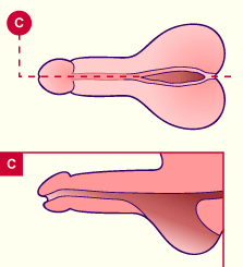
When no DHT is formed, no complete fusion of the genital swellings occurs, leading to scrotal hypospadia (opening of the urethra in the scrotal region). The ectopic testes are mostly normally developed and produce antimüllerian hormone (AMH; the paramesonephric duct is atrophied). The production of testosterone in puberty can lead to a further masculine development of the external genitals. Men with this disorder are also procreative! They have a male phenotype. |
|
Fig. 65 - Hypospadia of the scrotum |
|
|
|
|
|
Consequences of a testosterone deficiency:
All of the structures that depend on androgens for their development are affected; i.e., absent differentiation of the mesonephros (Wolff), missing descent of the testes, the external sexual organs and the behavior are feminine. Because the production of testosterone fails to occur in puberty, development of the secondary male sexual characteristics also does not occur.
|
|
|
|
Syndrome of the testicular feminization
Here a recessive disorder of the androgen receptors, connected with the X chromosome, is involved. The production of androgens is indeed normal, but the sensibility of the target organ/cells is damaged and the individual develops as if no testosterone is present. The testes are present and produce AMH, so that the paramesonephric duct atrophies. Due to missing androgen sensibility, though, the mesonephric tubules degenerate. Sometimes one finds a rudimentary vagina. The phenotype and also the psychic development are feminine.
The ectopic testes (either intra-abdominal, inguinal or located in the labia majora) secrete estrogen, leading to a mammary development in puberty.
The danger of a testicular tumor is elevated due to their ectopic position. Their early removal leads to a premature menopause and an absent development of the breasts. This measure is therefore controversial.
|
|
|
|
Pseudohermaphroditism in females
|
|
|
|
Under female pseudohermaphroditism one understands the existence of ambiguous external genitalia, even though ovaries and a 46,XX karyotype are present.
Ethiology:
The reason is an exposure of the fetus to androgens during the first trimester of pregnancy. The cause is a congenital suprarenal hyperplasia (recessive autosomal illness), a maternal suprarenal gland tumor, or a therapy of the pregnant mother with androgen hormones.
External manifestation:
The external sex organs differ in how strongly they are virilized; an enlarged clitoris with hypospadia is present; the scrotum is empty; and sometimes an obliterated vagina exists. The internal genitalia (ovary, fallopian tube and vagina) are normal and a pregnancy is possible! The virilization of the external genitalia can be complete at birth, but the testes are missing in the scrotum.
|
|
|
|
This consists of a developmental disorder of the ovaries with a feminine phenotype and a 45,X0 genotype or, in rare cases, a mosaic with genotype 45, X0 / 46, XX or 45, X0 / 46, XY, representing a male phenotype.
The external genitalia are normal and feminine, but the internal genitalia are incompletely developed with atrophied ovaries. At puberty the development of the breasts does not occur and the patients have a primary amenorrhoea. These women exhibit a stocky constitution (no spurt of growth during puberty) and a characteristic skin fold at the neck (pterygium colli). They are infertile, but the external genitalia are normal, making a normal sex life possible. Therapeutically, a hormone substitution is begun already in childhood.
|
|
|
|
This consists of a developmental disorder of the testes with a masculine phenotype and a 47, XXY genotype (80%) or, in 20%, a mosaic with 45, XY / 47, XXY genotypes. Certain genes, which influence the development of the testes, the production of sex hormones and growth, are localized on the X chromosome. There is, however, no uniform syndrome, but rather the definition depends on the genotype of the individual.
Klinefelter's syndrome is then characterized by the following symptoms: sterility (the seminiferous tubules are hyalinized), incomplete virilization with a testicular hypoplasia, small penis, large body growth and partial gynecomastia, feminine hair type at the beginning of puberty and finally osteoporosis and mental retardation with behavioral problems.
|
|
|
|
|