21.6 Pathology


Abnormalities of female genitalia

Quiz

Quiz 17


A disturbed fusion of the lower section of the paramesonephric duct (Müller) can lead to a variety of abnormalities in the utero-vaginal region. Such abnormalities in the genital region are almost always associated with such of the urinary tract, since these two systems are closely connected with each other.


Utero-vaginal abnormalities


As we saw earlier, the absence of AMH leads without fail to a further development of the paramesonephric duct. Three phases can be distinguished:

  • The spread of the paramesonephric duct down to the urogenital sinus.
  • The fusion on both sides of the lower third of the paramesonephric duct out of which the uterus and the upper 3/4 of the vagina arise
  • The resorption of the dividing wall of the paramesonephric duct on both sides after fusion (end of the 3rd month)


An absent or incomplete migration of the paramesonephric duct in the direction of the SUG is responsible for an atresia and/or complete or incomplete aplasia of the uterus, which is usually associated with renal abnormalities. This syndrome is called the Maye Rokitansky Kuster Hauser syndrome. Fig. 75 - Unilateral atresia, leading to
a uterus unicornis unicollis


A partial or complete failure of the lower parts of the two paramesonephric ducts (Müller) to fuse or an incomplete development (atresia) of one of two paramesonephric ducts is responsible for the formation of a uterus bicornis uni- or bicollis with or without doubling of the vagina. The uterus bicornis unicollis is encountered the most frequently. Fig. 76 - Uterus didelphys bicollis


Fig. 77 - Uterus bicornis bicollis Fig. 78 - Uterus bicornis unicollis


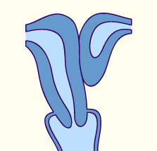
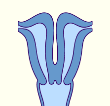
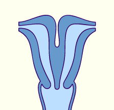
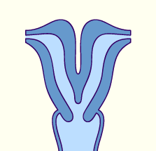
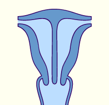
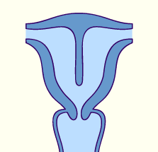
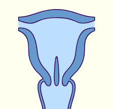
The absent resorption of the median dividing wall of the two paramesonephric ducts (Müller) leads to a septated uterus:
  • Uterus septus (from the body to the uterine cervix)
  • Uterus subseptus (only in the body region)
  • Uterus subseptus (only in the cervical region)

When no vaginal plate develops, this leads to a vaginal aplasia that, though, only very rarely occurs in isolation. Due to their partly common origin uterine abnormalities are mostly associated with those of the vagina.

Fig. 79 - Uterus septus


Fig. 80 - Uterus septus subtotalis unicollis Fig. 81 - Uterus septus unicornis bicollis


Previous page | Next page