21.6 Pathology


Inguinal hernia / hydrocele / cysts


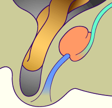
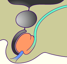
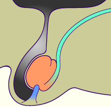
Normally, during the first year of life the upper section of the vaginal process closes and only the peritoneo-vaginal ligament remains. Only a small sack on the ventral surface of the testes remains. A double-walled serous structure, the vaginal tunica, is involved.

If the vaginal process is not obliterated, but stays open, intestinal loops can slide into it and cause a congenital inguinal hernia. Such hernias can be more or less extensive, depending on the dehiscence between the visceral peritoneum of the intestinal loops and the vaginal process (parietal peritoneum).

Quiz

Quiz 30


Fig. 71 - Incomplete inguinal hernia Fig. 72 - Complete inguinal hernia


It can also happen that the opening is too small for the intestinal loop and instead peritoneal fluid can collect therein, forming a cyst in the spermatic cord, if the fluid is closed in above the testes or a testicular hydrocele can be formed when a serous discharge collects between the layers of the vaginal tunica.


Fig. 73 - Cyst of the spermatic cord Fig. 74 - Hydrocele


Testicular torsion


Torsion of the spermatic cord, mistakenly called a testicular torsion, is connected with a congenital anomaly of the testicular fixation at the lower scrotal pole. Thereby the spermatic cord (deferent duct and testicular blood vessels) can twist around its axis.
Through this, the testicular artery gets compressed and an ischemia (insufficient perfusion of the testis) arises. This is always a urologic emergency. The treatment is always surgical and must take place within the first 6 hours to avoid irreversible damage to the testis. This torsion can appear at any time up to adolescence, but is rarely encountered in adults.


Previous page | Next page