|

|
|
|
7.2 The trilaminar germ disk (3rd week)
|
|
|
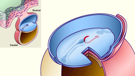
| Fig. 5 - Primitive streak seen dorsally |
|
Legend |
|
|
Fig. 5
The red arrow shows schematically the migration direction of the epiblast cells coming from the primitive node to form the notochordal process and later the notochord.
|
Fig. 5 a - The chordal process at
roughly the 19th day (Stage 7) |
|
Fig. 5 b - The chordal process at
roughly the 19th day (Stage 7) |
|
Legend |
1
3
|
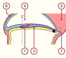
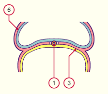
Chordal process
Embryonic endoblast |
|
|
|
4
5
6
7 |
Amniotic cavity
Body stalk
Extraembryonic mesoblast
Allantois |
|
|
|
Fig. 5a
Schematic diagram showing the genesis of the chordal process at the 19th day through the invagination of epiblast cells coming from the primitive node.
Fig. 5b
Cut along C.
|
Fig. 5 c - The chordal process at
roughly the 21rst day (Stage 7) |
|
Fig. 5 d - The chordal process at
roughly the 21rst day (Stage 7) |
|
Legend |
1
2
3
4 |
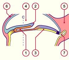
Chordal process
Primitive node
Embryonic endoblast
Amniotic cavity |
|
|
|
5
6
7 |
Body stalk
Extraembryonic mesoblast
Allantois |
|
|
|
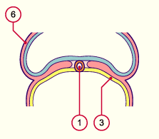
Fig. 5c
Schematic diagram showing the genesis of the axial canal through invagination of epiblast cells that migrate into the region of the primitive node (on roughly the 21rst day).
Fig. 5d
Cut along C
|
Fig. 6a - The chordal process at
roughly the 23rd day (Stage 8) |
|
Fig. 6b - The chordal process at
roughly the 23rd day (Stage 8) |
|
Legend |
1
2
3
4
5
6
|
Fused chordal process
Prechordal plate
Pharyngeal membrane
Embryonic endoblast
Amniotic cavity
Neural groove |
|
|
|
7
8
9
10
11
|
Canalis neurentericus
Intraembryonic mesoblast
Cloacal membrane
Umbilical vesicle
Allantois |
|
|
|
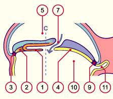
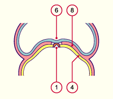
Fig. 6a
The notochord has separated itself from the endoblast. At the caudal end of the notochord, in the region of the primitive node, thanks to the neurenteric canal, a temporary communication takes place between the umbilical vesicle and the amniotic cavity (arrow).
Fig. 6b
Cut along C
|
|
On the 25th day  9 9 the chordal plate cuts itself off from the entoderm, which then fuses again, and forms a complete cord: the notochord. This is in the middle of the mesoderm, between the ectodern and endoderm, and plays a role in the induction of the neuroectoblast that lies over it. Moreover, the notochord plays a role in the genesis of the vertebral body and becomes the nucleus pulposus in the intervertebral disk. the chordal plate cuts itself off from the entoderm, which then fuses again, and forms a complete cord: the notochord. This is in the middle of the mesoderm, between the ectodern and endoderm, and plays a role in the induction of the neuroectoblast that lies over it. Moreover, the notochord plays a role in the genesis of the vertebral body and becomes the nucleus pulposus in the intervertebral disk.
|
|
|
Fig. 7a - The chordal process at
roughly the 25th day (Stage 9) |
|
Fig. 7b - The chordal process at
roughly the 25th day (Stage 9) |
|
Legend |
1
2
3
4
5
6
7 |
Chordal process
Embryonic endoderm
Amniotic cavity
Neural groove
Body stalk
Intraembryonic mesoblast
Prechordal plate |
|
|
|
8
9
10
11
12
13
|
Pharyngeal membrane
Cloacal membrane
Aortae
Umbilical veins
Cardiogenic plate
Allantois |
|
|
|
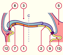
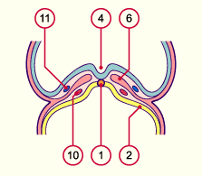
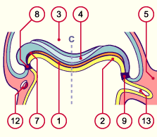
Fig. 7a
At around the 20th day the chordal tissue bonds with the endoderm and thus forms the chordal plate.
Between the 22nd and 24th day it cuts itself off from the endoderm (the endoderm fuses again) in order to form a complete cord: the notochord. This finds itself in the middle of the mesoderm, between the ectoderm and endoderm.
Fig. 7b
Cut along C
|
Fig. 7c - The chordal process at
roughly the 28th day (Stage 10) |
|
Fig. 7d - The chordal process at
roughly the 28th day (Stage 10) |
|
Legend |
1
2
3
4
5
6
7
|
Chordal process
Embryonic endoblast
Amniotic cavity
Neural tube
Body stalk
Intraembryonic mesoblast
Prechordal plate |
|
|
|
8
9
10
11
12
13
|
Pharyngeal membrane
Cloacal membrane
Aortae
Umbilical veins
Cardiogenic plate
Allantois |
|
|
|
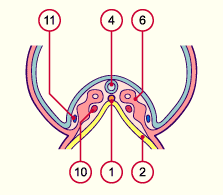
Fig. 7c
The endoderm, under the notochord, comes together again. At this point the neural groove has partly fused into becoming a neural tube.
Fig. 7d
Cut along C
|
Summary:
the notochord determines the longitudinal axis of the embryo. It defines the future situation of the vertebral body and induces the ectoblast in its differentiation to become the neural plate. |
|
|
|
|
More info
|
|
In the literature there is some disagreement related to the delimitation and fate of the prechordal plate.
|
|
|
|
|

