|

|
|
|
7.2 The trilaminar germ disk (3rd week)
|
|
|
|
The paraxial mesoblast and the differentiation of the somites
|
|
|
|
The paraxial mesoblast comes from the epiblast cells that migrated into the region of the primitive node or the cranial portion of the primitive streak. It forms a pair of cylinder-shaped epithelially-organized mesenchyma segments that are in the immediate vicinity of the neural tube and the notochord.
After the beginning of the 3rd week, these cylinders become segmented from the cranial to the caudal end into so-called somitomeres (process of metamerization). Originally, each consists of a pseudostratified epithelium that is arranged around a central cavity, the somitocoel.
|
|
|
More info
|
|
The metameres are based on the division of the original embryo into several segments.
|
|
|
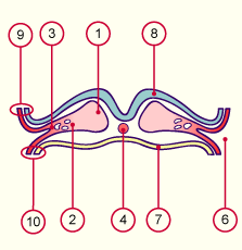
| Fig. 15 - Genesis of the intraembryonic coelom, at roughly the 23rd day |
|
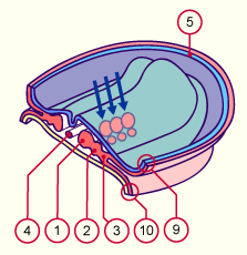
Fig. 16 - Appearance of the somitomeres, at roughly the 25th day |
|
Legend |

1
2
3
4
5
6
|
Paraxial mesoblast
Intermediate mesoblast
Lateral plate mesoblast
Chordal process
Sectional edge of the amnion
Intraembryonic coelom |
|
|
|

7
8
9
10
|
Endoblast
Ectoblast
Somatopleure with ectoblast
Splanchnopleure with endoblast |
|
|
|
Fig. 15
Transversal section through a 23-day-old embryo. One can recognize the first space of the future intraembryonic coelom.
Fig. 16
Transverse section with a dorsal view at around the 25th day. The cylindrical cell collections of the paraxial mesoderm segment form the somitomeres (arrows). The intermediate mesoblast is involved in the urogenital development.
|
|
More info
|
|
The somites are embryonic transitional organs that are formed through the segmentation of the paraxial mesenchyma. They organize themselves without cell differentiation (primary organs). They are responsible for the segmental organization of the embryo and contribute to its restructuring.They contain the precursor cells for the axial skeleton (sclerotome), the striated musculature of the neck, the trunk and the extremities (myotome), as well as those of the subcutaneous tissue and skin (dermatome). The somites are the prerequisites for metamerism. The segmental (metamere) partitioning of the spine, the neural tube, the trunk wall and the thorax (ribs) depends on the ordered arrangement of the somites.
|
|
|
|
|
|
|

